 Back
BackDigestive System: Structure, Function, and Physiology
Study Guide - Smart Notes
 Tailored notes based on your materials, expanded with key definitions, examples, and context.
Digestive System Overview
Major Functions and Organization
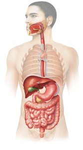
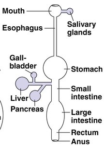
The digestive system is responsible for breaking down food into nutrients, which are then absorbed into the bloodstream and delivered to cells throughout the body. It consists of the gastrointestinal (GI) tract—a continuous tube with two openings—and accessory organs that aid in digestion.
GI Tract (Alimentary Canal): Includes the mouth, esophagus, stomach, small intestine, and large intestine.
Accessory Structures: Salivary glands, tongue, teeth, gall bladder, liver, and pancreas (not part of the GI tract but essential for digestive processes).
Main Functions: Ingestion, secretion, propulsion (e.g., peristalsis), digestion (mechanical and chemical), absorption, and defecation (elimination).
Digestive Processes
Ingestion: Taking in food via the mouth.
Secretion: Release of water, acid, buffers, and enzymes into the lumen of the GI tract.
Propulsion: Movement of food through the tract, primarily by peristalsis.
Digestion:
Mechanical: Physical breakdown (e.g., mastication, churning, segmentation).
Chemical: Enzymatic hydrolysis of macromolecules.
Absorption: Passage of digested nutrients into blood or lymph.
Defecation: Elimination of indigestible substances as feces.
Histological Structure of the GI Tract
Four Layers of the Alimentary Canal Wall
The wall of the GI tract is composed of four main layers, each with distinct structures and functions:
Mucosa: Innermost layer; consists of epithelium, lamina propria, and muscularis mucosae. Functions in secretion and absorption.
Submucosa: Connective tissue containing blood vessels, nerves, and glands.
Muscularis: Two to three layers of smooth muscle responsible for peristalsis and segmentation.
Serosa/Adventitia: Outermost layer; serosa is present within the peritoneal cavity, while adventitia is found outside it.
Peritoneum and Mesenteries
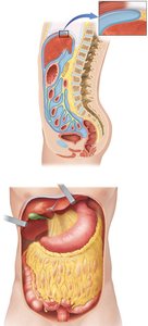
Peritoneum: Serous membrane lining the abdominopelvic cavity.
Parietal Peritoneum: Lines the inner wall of the cavity.
Visceral Peritoneum: Covers the organs and is continuous with the serosa.
Functions: Protection, suspension of organs, and providing a route for blood vessels and nerves.
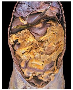
Major Folds: Mesenteries, greater omentum, and lesser omentum.
Oral Cavity, Teeth, and Salivary Glands
Oral Cavity Structure and Function
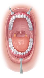
The oral cavity (mouth) is formed by the cheeks, lips, tongue, and hard and soft palates. It contains the teeth and salivary glands and is the site where ingestion, secretion, mixing/propulsion, and chemical digestion begin.
Teeth: Structure and Types
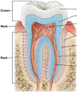
Regions: Crown, neck, and root.
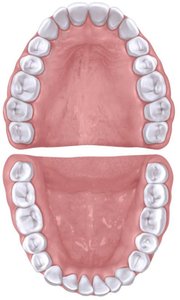
Dentitions: Primary (deciduous) and secondary (permanent).
Types: Incisors (cutting), canines (shearing), premolars and molars (crushing and grinding).
Function: Mastication increases the surface area of food, enhancing enzymatic activity.
Salivary Glands
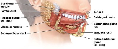
Three Paired Glands: Parotid, submandibular, and sublingual glands.
Saliva Composition: 99.5% water, 0.5% solutes (enzymes, salts, organic molecules, dissolved gases).
Functions: Lubricates and moistens food; enzymes initiate chemical breakdown.
Pharynx and Esophagus
Pharynx
Structure: Connects the oral cavity to the esophagus; composed of three regions (upper, middle, lower) with varying muscle types (skeletal to smooth).
Function: Propulsion of food; secretes mucus and transports food to the stomach.
Esophagus
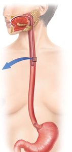
Muscle Composition: Upper third (skeletal muscle), middle third (mixed), lower third (smooth muscle).
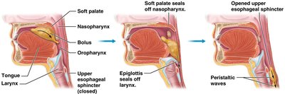
Sphincters: Upper and lower esophageal sphincters regulate passage of food.
Deglutition (Swallowing)
Stages:
Voluntary: Tongue pushes food to oropharynx.
Pharyngeal: Epiglottis closes off the airway; food moves to esophagus.
Esophageal: Peristalsis moves food through the esophagus to the stomach.
Stomach
Structure and Function
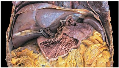
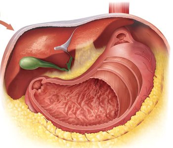
Shape: J-shaped organ bounded by esophageal and pyloric sphincters.
Regions: Cardia, fundus, body, and pylorus.
Muscle Layers: Three layers of smooth muscle for mixing and churning.
Rugae: Folds in the mucosa when the stomach is empty.
Functions: Mixing, holding area for food, some absorption (e.g., alcohol, aspirin), and chemical digestion.
Histology of the Stomach
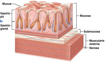
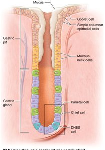
Mucosa: Simple columnar epithelium, gastric pits, and gastric glands.
Gastric Pits: Contain mucous cells (secrete mucus) and mucous neck cells (secrete acidic mucus).
Gastric Glands:
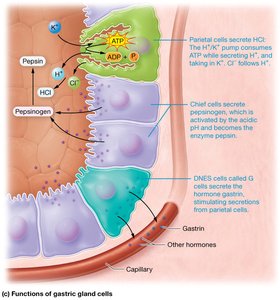
Parietal cells: Secrete H+, Cl-, and intrinsic factor (for vitamin B12 absorption).
Chief cells: Secrete pepsinogen (activated to pepsin by HCl) and gastric lipase.
Enteroendocrine cells (G cells): Secrete gastrin (hormone).

Stomach Digestion and Regulation
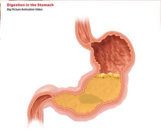
Mechanical Digestion: Peristaltic contractions mix food with gastric juices.
Chemical Digestion:
Pepsin: Digests proteins in the presence of HCl.
Gastric lipase: Digests some lipids.
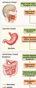
Phases of Digestion:
Cephalic Phase: Initiated by sensory input (sight, smell, taste, thought of food); stimulates gastric secretion and motility.
Gastric Phase: Triggered by food in the stomach; increases secretion and motility.
Intestinal Phase: Begins as chyme enters the small intestine; regulates further gastric activity.
Small Intestine
Structure and Regions
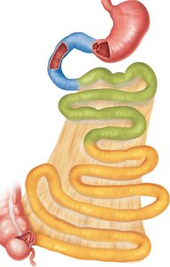
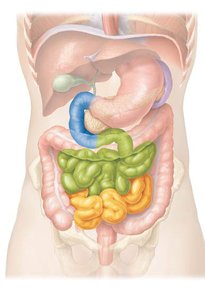
Primary Site: Chemical digestion and nutrient absorption.
Regions: Duodenum, jejunum, ileum.
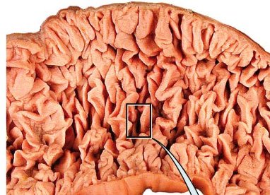
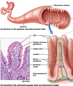
Specializations: Circular folds (plicae circulares) increase surface area for absorption.
Histology and Absorptive Structures
Villi: Fingerlike projections of the mucosa; each contains a lacteal (lymphatic capillary) and blood supply.
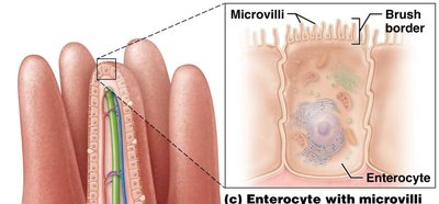
Microvilli: Extensions of enterocyte membranes forming the brush border; contain enzymes for final digestion steps.
Intestinal Glands: Invaginations in the mucosa; secrete intestinal juices.
Submucosal Glands: Especially in the duodenum; secrete alkaline mucus to neutralize chyme.
MALT (Peyer’s patches): Lymphoid tissue in the ileum for immune defense.
Cell Types in the Small Intestine
Absorptive Cells (Enterocytes): Apical surface with microvilli; absorb nutrients.
Goblet Cells: Secrete mucus for lubrication and protection.
Enteroendocrine Cells: Secrete hormones (secretin, CCK, GIP) that regulate digestive activity.
Motility in the Small Intestine
Segmentation: Circular muscle contractions mix chyme and increase contact with absorptive surfaces.
Peristalsis: Alternating contractions of circular and longitudinal muscles propel contents forward.
Large Intestine
Structure and Function
Functions: Completes absorption of water, produces certain vitamins, forms and expels feces.
Regions: Cecum (with appendix), colon (ascending, transverse, descending, sigmoid), rectum, anal canal.
Histology
Mucosa: Increased goblet cells; no villi or circular folds.
Muscularis: Longitudinal muscle forms taeniae coli, creating pouches called haustra.
Defecation Reflex
Triggered by stretch receptors in the sigmoid colon.
Involves voluntary contraction of the diaphragm and abdominal muscles, and relaxation of the external anal sphincter.
Accessory Organs: Pancreas, Liver, and Gallbladder
Pancreas
Location: Posterior to the stomach.
Structure: 1% endocrine (islets of Langerhans), 99% exocrine (acini).
Digestive Enzymes:
Pancreatic amylase (starch digestion)
Trypsin, chymotrypsin, carboxypeptidase (protein digestion)
Pancreatic lipase (fat digestion)
Ribonuclease, deoxyribonuclease (nucleic acid digestion)
Liver and Gallbladder
Liver: Second largest organ; produces bile for fat emulsification, processes nutrients, detoxifies substances, and stores glycogen.
Gallbladder: Stores and releases bile produced by the liver.
Bile Ducts: Common bile and pancreatic ducts empty into the duodenum at the duodenal papilla.
Liver Histology and Blood Supply
Hepatocytes: Arranged in lobules around a central vein.
Portal Triads: Located between lobules; contain a bile duct, hepatic arteriole (O2-rich blood), and hepatic portal venule (nutrient-rich, O2-poor blood).
Blood drains into hepatic sinusoids and then to the central vein.
Nutrient Digestion and Absorption
Enzymatic Hydrolysis and Chemical Digestion
Carbohydrates: Digested into monosaccharides (e.g., glucose, galactose).
Proteins: Digested into amino acids.
Lipids: Digested into fatty acids and monoglycerides.
Lipid Digestion and Absorption
Lipids are broken apart by stomach churning and gastric lipase.
In the small intestine, bile salts emulsify lipids.
Pancreatic lipase digests lipids into free fatty acids and monoglycerides.
Bile salts form micelles with digested lipids, facilitating absorption.
Absorbed lipids are reassembled into chylomicrons and enter lacteals (lymphatic capillaries), bypassing the liver initially.
Absorption of Water, Electrolytes, and Vitamins
Most water (>8L/day) is absorbed in the small intestine; the remainder in the large intestine.
Vitamins:
Water-soluble (B, C): Absorbed in the small intestine (except B12, which requires intrinsic factor and is absorbed in the ileum).
Fat-soluble (A, D, E, K): Packaged into micelles and absorbed with lipids.
Summary Table: Structure and Function of Digestive Organs
Organ | Main Function(s) | Key Features |
|---|---|---|
Mouth | Ingestion, mechanical and chemical digestion | Teeth, tongue, salivary glands |
Esophagus | Propulsion | Peristalsis, sphincters |
Stomach | Mixing, chemical digestion, some absorption | Rugae, gastric glands, three muscle layers |
Small Intestine | Chemical digestion, nutrient absorption | Villi, microvilli, circular folds |
Large Intestine | Water absorption, feces formation | Haustra, taeniae coli, goblet cells |
Liver | Bile production, metabolism | Lobules, portal triads |
Gallbladder | Bile storage and release | Muscular sac |
Pancreas | Secretion of digestive enzymes | Acini, islets of Langerhans |
