 Back
BackDigestive System: Structure, Function, and Physiology
Study Guide - Smart Notes
 Tailored notes based on your materials, expanded with key definitions, examples, and context.
The Digestive System: Overview
Main Functions of the Digestive System
The digestive system is responsible for the intake, breakdown, absorption, and elimination of food. Its primary functions include:
Ingestion: Taking in food through the mouth.
Mechanical and Chemical Breakdown: Physically and chemically breaking down food into smaller molecules.
Absorption: Transporting digested nutrients into the bloodstream or lymphatic system.
Elimination: Removing indigestible substances from the body as feces.
Organization of the Digestive System
Alimentary Canal and Accessory Organs
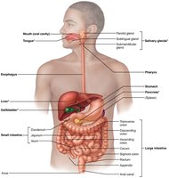
The digestive system consists of the alimentary canal (gastrointestinal tract) and accessory digestive organs.
Alimentary Canal: A continuous muscular tube from mouth to anus, including the mouth, pharynx, esophagus, stomach, small intestine, large intestine, and anus.
Accessory Organs: Teeth, tongue, gallbladder, salivary glands, liver, and pancreas. These organs assist in digestion by producing secretions or aiding in mechanical breakdown.
Digestive Processes
Six Essential Activities
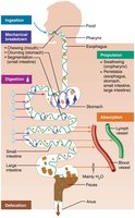
Food processing in the digestive system involves six key activities:
Ingestion: Eating food.
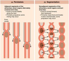
Propulsion: Moving food through the alimentary canal, including swallowing and peristalsis (alternating waves of contraction and relaxation).
Mechanical Breakdown: Chewing, mixing, churning, and segmentation (local constriction that mixes food with digestive juices).
Digestion: Enzymatic breakdown of complex food molecules into chemical building blocks.
Absorption: Passage of digested fragments from the GI tract into blood or lymph.
Defecation: Elimination of indigestible substances as feces.
Relationship to the Peritoneum
Peritoneum and Mesenteries
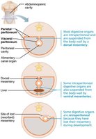
The peritoneum is a serous membrane lining the abdominal cavity, consisting of:
Visceral Peritoneum: Covers external surfaces of most digestive organs.
Parietal Peritoneum: Lines the body wall.
Peritoneal Cavity: Fluid-filled space between the two peritoneums, lubricating mobile organs.
Mesentery: Double layer of peritoneum that provides routes for blood vessels, lymphatics, and nerves, holds organs in place, and stores fat.
Intraperitoneal Organs: Located within the peritoneum.
Retroperitoneal Organs: Located outside or posterior to the peritoneum (e.g., most of the pancreas, duodenum, parts of the large intestine).
Histology of the Alimentary Canal
Four Basic Layers (Tunics)
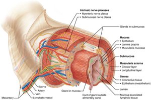
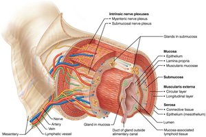
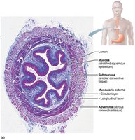
All digestive organs share a common structural plan with four layers:
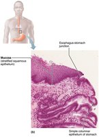
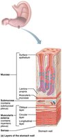
Mucosa: Innermost layer; secretes mucus, digestive enzymes, and hormones; absorbs end products; protects against infection. Composed of epithelium, lamina propria, and muscularis mucosae.
Submucosa: Areolar connective tissue with blood and lymphatic vessels, lymphoid follicles, and submucosal nerve plexus.
Muscularis Externa: Responsible for segmentation and peristalsis; consists of inner circular and outer longitudinal muscle layers. Circular layer forms sphincters in some regions.
Serosa: Outermost layer; visceral peritoneum. In the esophagus, replaced by fibrous adventitia.
Control of Digestive System
Enteric Nervous System
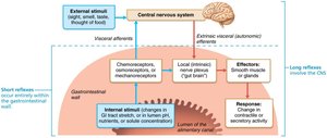
The GI tract has its own nervous system, the enteric nervous system (ENS), also called the "gut brain." It contains more neurons than the spinal cord and controls GI tract motility.
Submucosal Nerve Plexus: Regulates glands and smooth muscle in the mucosa.
Myenteric Nerve Plexus: Controls GI tract motility.
Short Reflexes: Mediated by ENS, respond to stimuli in the GI tract.
Long Reflexes: Involve the autonomic nervous system, responding to stimuli inside or outside the gut.
Parasympathetic: Enhances digestive activity; Sympathetic: Inhibits digestion.
Mouth and Associated Organs
Oral Cavity Structure
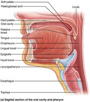
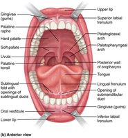
The mouth (oral cavity) is the entry point for food and is lined with stratified squamous epithelium for protection. Key structures include:
Lips and Cheeks: Contain muscles for manipulating food.
Palate: Hard and soft regions form the roof of the mouth.
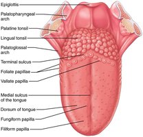
Tongue: Skeletal muscle that mixes food, forms the bolus, and initiates swallowing and speech.
Tongue and Papillae
The tongue is covered with papillae, which aid in taste and food manipulation:
Filiform Papillae: Provide friction, no taste buds.
Fungiform, Vallate, and Foliate Papillae: Contain taste buds.
Salivary Glands
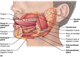
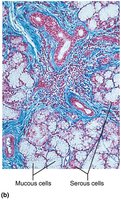
Salivary glands produce saliva, which cleanses the mouth, dissolves food chemicals, moistens food, and begins starch digestion with amylase. Major glands include:
Parotid: Anterior to ear, opens near upper molar.
Submandibular: Medial to mandible, opens at base of lingual frenulum.
Sublingual: Under tongue, opens via multiple ducts.
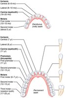
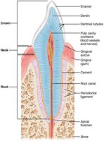
Teeth
Teeth are essential for mastication (chewing), breaking food into smaller fragments. Types include:
Incisors: Cutting.
Canines: Tearing.
Premolars and Molars: Grinding and crushing.
Pharynx and Esophagus
Structure and Function
The pharynx and esophagus serve as passageways for food from the mouth to the stomach. The esophagus is a muscular tube with all four alimentary canal layers and transitions from skeletal to smooth muscle along its length.
Deglutition (Swallowing)
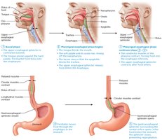
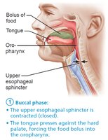
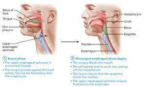
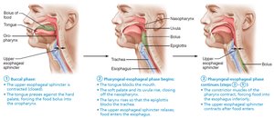
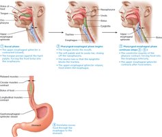
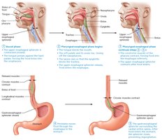
Swallowing involves voluntary and involuntary phases, coordinated by the medulla and lower pons. The process ensures food is safely transported from the mouth to the stomach.
The Stomach
Gross Anatomy
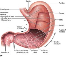
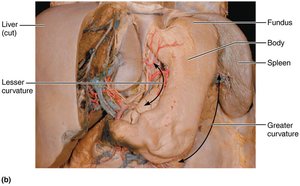
The stomach is a muscular, expandable organ that stores food and begins protein digestion. It is divided into regions: cardia, fundus, body, and pyloric part. The stomach wall has three muscle layers for churning and mixing food.
Microscopic Anatomy
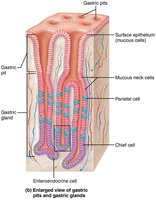
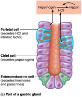
The stomach mucosa contains gastric pits leading to gastric glands, which produce gastric juice. Key cell types include:
Mucous Neck Cells: Secrete acidic mucus.
Parietal Cells: Secrete HCl and intrinsic factor.
Chief Cells: Secrete pepsinogen and lipases.
Enteroendocrine Cells: Secrete hormones and paracrines.
Gastric Secretion and Regulation
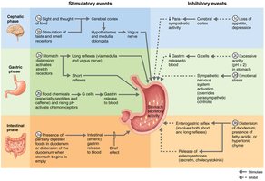
Gastric secretion is regulated by neural (vagus nerve) and hormonal (gastrin) mechanisms, occurring in three phases: cephalic, gastric, and intestinal. Gastric juice contains HCl, pepsin, and intrinsic factor, essential for protein digestion and vitamin B12 absorption.
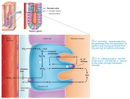
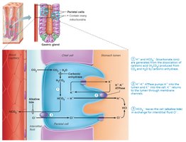
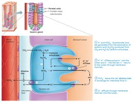
Mechanism of HCl Formation
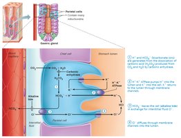
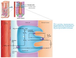
Parietal cells secrete HCl by pumping H+ into the stomach lumen via H+/K+ ATPase, while Cl− is transported to combine with H+ to form HCl. The process also results in an alkaline tide (increased HCO3− in blood).
Small and Large Intestine
Small Intestine
The small intestine is the major site for digestion and absorption. It is divided into the duodenum, jejunum, and ileum. Structural modifications (circular folds, villi, microvilli) increase surface area for absorption.
Large Intestine
The large intestine absorbs water and electrolytes, forming and storing feces. It includes the cecum, colon, rectum, and anal canal. Unique features include teniae coli, haustra, and epiploic appendages.
Digestive Physiology: Digestion and Absorption
Carbohydrate Digestion and Absorption
Carbohydrates are broken down by amylases and brush border enzymes into monosaccharides, which are absorbed via secondary active transport (with Na+) and facilitated diffusion.
Protein Digestion and Absorption
Proteins are digested by pepsin in the stomach and by pancreatic and brush border enzymes in the small intestine. Amino acids are absorbed via secondary active transport and facilitated diffusion.
Lipid Digestion and Absorption
Lipids are emulsified by bile salts and digested by pancreatic lipases. Products are absorbed as micelles, reassembled into triglycerides, and transported as chylomicrons into lymphatics.
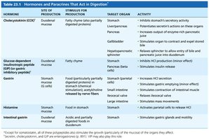
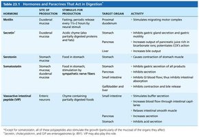
Summary Table: Hormones and Paracrines in Digestion
Hormone | Site of Production | Target Organ | Activity |
|---|---|---|---|
Cholecystokinin (CCK) | Duodenum | Stomach, pancreas, gallbladder | Inhibits gastric secretion, stimulates enzyme-rich pancreatic juice, stimulates gallbladder contraction |
Secretin | Duodenum | Stomach, pancreas, liver | Inhibits gastric secretion, stimulates bicarbonate-rich pancreatic juice, increases bile output |
Gastrin | Stomach | Stomach, small intestine, ileocecal valve, large intestine | Stimulates HCl secretion, stimulates gastric emptying, stimulates contraction of intestinal muscle |
Motilin | Duodenum | Proximal duodenum | Stimulates migrating motor complex |
Key Equations
HCl Formation in Parietal Cells:
is pumped into the stomach lumen via H+/K+ ATPase, while is exchanged for , which then combines with to form HCl.
