Back
BackDigestive System: Structure, Function, and Processes
Study Guide - Smart Notes
 Tailored notes based on your materials, expanded with key definitions, examples, and context.
Digestive System Overview
General Anatomical Features
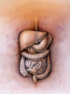
The digestive system is responsible for providing a steady supply of nutrients, water, and inorganic ions to the body. It consists of the digestive tract (alimentary canal) and accessory structures. The tract includes the mouth, pharynx, esophagus, stomach, small intestine, and large intestine. Accessory structures such as teeth, tongue, salivary glands, pancreas, liver, and gallbladder assist in mechanical digestion or secrete substances that aid digestion.
Ingestion: Taking food into the mouth.
Propulsion: Moving food through the tract, mainly by swallowing and peristalsis.
Mechanical Breakdown: Physical breakdown of food (chewing, churning, segmentation).
Digestion: Chemical breakdown of food into absorbable molecules.
Absorption: Transport of digested nutrients into blood or lymph.
Defecation: Elimination of indigestible substances as feces.
Motility Patterns
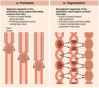
Two primary types of motility patterns move and mix food along the digestive tract:
Peristalsis: Adjacent segments of the alimentary canal organs alternately contract and relax, propelling food distally.
Segmentation: Nonadjacent segments contract and relax, mixing food and breaking it down mechanically; some propulsion may occur.
Peritoneum and Mesenteries
Peritoneum
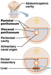
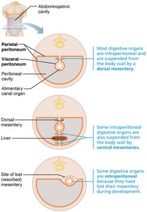
The peritoneum is a double-layered serous membrane associated with the abdominal organs. The visceral peritoneum covers the organs, while the parietal peritoneum lines the abdominopelvic cavity. The space between them is the peritoneal cavity, filled with serous fluid to reduce friction.
Mesenteries
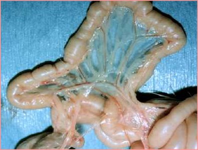
A mesentery is a double layer of peritoneum that extends from the body wall to digestive organs. It allows blood vessels, lymphatics, and nerves to reach the organs, holds organs in place, and stores fat. Most mesenteries are dorsal, but some organs (e.g., pancreas, duodenum) are retroperitoneal because they lost their mesentery during development.
Blood Supply of the Digestive System
Splanchnic Circulation
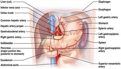
The digestive system requires an extensive blood supply. Splanchnic circulation includes arteries branching from the aorta (hepatic, splenic, left gastric, inferior and superior mesenteric arteries) and the hepatic portal circulation, which delivers nutrient-rich blood from digestive organs to the liver for processing. About one-quarter of cardiac output goes to the digestive system, increasing after meals.
Histology of the Alimentary Canal
Four Basic Layers
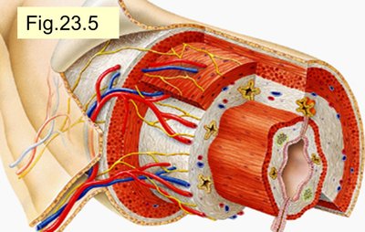
The wall of the alimentary canal consists of four layers:
Mucosa: Innermost layer; secretes mucus, enzymes, and hormones; absorbs nutrients; protects against infection.
Submucosa: Connective tissue with blood vessels, lymphatics, nerves, and glands.
Muscularis externa: Responsible for segmentation and peristalsis; usually has inner circular and outer longitudinal layers (stomach also has an oblique layer).
Serosa: Outermost layer; visceral peritoneum. In the esophagus, this is replaced by adventitia.
Mucosa Details
Epithelium: Stratified squamous (mouth, esophagus, anus) or simple columnar (stomach, intestines) with goblet cells (produce mucus).
Lamina propria: Connective tissue with capillaries and MALT (mucosa-associated lymphoid tissue).
Muscularis mucosae: Thin layer of smooth muscle for local movements.
Submucosa and Muscularis Externa
Submucosa: Contains exocrine glands and the submucosal plexus (regulates glands and muscle).
Muscularis externa: Contains the myenteric plexus (controls GI tract motility) and forms sphincters.
Neural Control of the Digestive System
Enteric Nervous System
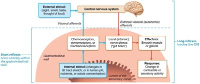
The enteric nervous system (ENS) consists of the submucosal and myenteric plexuses. It regulates GI tract activity through short (local) and long (CNS-mediated) reflexes, responding to stimuli such as stretch, pH, and chemical composition.
The Mouth and Associated Structures
Anatomy of the Oral Cavity
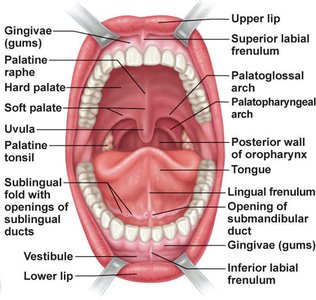
The mouth includes the vestibule, oral cavity proper, and oropharynx. The walls are lined with stratified squamous epithelium, with keratinization in areas subject to abrasion. The lips and cheeks contain skeletal muscle (orbicularis oris and buccinators) to hold food during chewing and aid in speech. The hard and soft palates form the roof, with the soft palate rising during swallowing to close off the nasopharynx. The tongue, with intrinsic and extrinsic muscles, positions and mixes food, forming a bolus for swallowing.
Tongue and Papillae
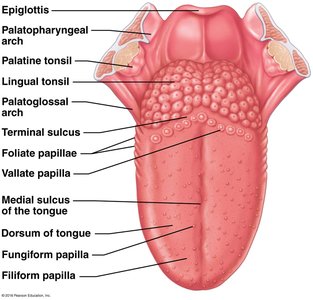
The tongue's surface contains four types of papillae:
Filiform: Conical, keratinized, provide friction (no taste buds).
Fungiform: Mushroom-shaped, scattered, contain taste buds.
Vallate (circumvallate): Large, V-shaped row at back, contain taste buds.
Foliate: On sides, contain taste buds (especially in children).
Salivary Glands and Saliva
Three pairs of extrinsic salivary glands (parotid, submandibular, sublingual) and small intrinsic buccal glands secrete saliva. Saliva cleanses the mouth, dissolves food for taste, moistens and compacts food, and begins starch digestion (amylase). It is mostly water, slightly acidic, and contains electrolytes, enzymes, proteins (mucin, lysozyme, IgA), and metabolic wastes. Salivation is controlled by the parasympathetic nervous system and can be stimulated by food, smell, or thought of food.
Teeth and Mastication
Dentition and Dental Formula
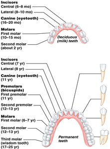
Humans have two sets of teeth: primary (deciduous) and permanent. Teeth are classified by shape and function: incisors (cutting), canines (tearing), premolars (grinding/crushing), and molars (grinding). The dental formula indicates the number and position of teeth in each quadrant of the mouth.
Tooth Structure
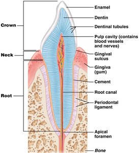
Each tooth has a crown (above gum, covered by enamel), neck, and root (embedded in jawbone). Enamel is the hardest substance in the body but is avascular and non-regenerative. Dentin lies beneath enamel and is maintained by odontoblasts. The root is covered by cement, which attaches the tooth to the periodontal ligament. The pulp cavity contains blood vessels and nerves.
Mastication (Chewing)
Mastication is the process of chewing, involving coordinated action of the jaws, lips, cheeks, and tongue. It is partly voluntary but largely reflexive, breaking food into smaller pieces and mixing it with saliva to form a bolus.
Swallowing and the Esophagus
Deglutition (Swallowing)
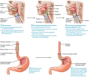
Swallowing involves moving food from the mouth to the stomach and consists of two phases:
Buccal phase: Voluntary; tongue pushes bolus into pharynx.
Pharyngeal-esophageal phase: Involuntary; coordinated by the swallowing center in the medulla and lower pons, involving closure of respiratory passages and peristalsis in the esophagus.
Esophagus Structure
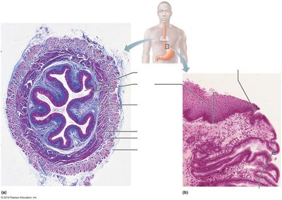
The esophagus is a muscular tube (~25 cm) that connects the pharynx to the stomach. Its upper third is skeletal muscle, transitioning to smooth muscle in the lower two-thirds. The mucosa and submucosa are folded when empty and flatten during food transit. The esophagus has an adventitia instead of a serosa.
Stomach: Structure and Function
Gross Anatomy
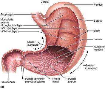
The stomach acts as a temporary storage tank, initiates protein digestion, and converts food to chyme. It has four regions: cardia, fundus, body, and pyloric region. The stomach wall contains rugae (folds) that allow expansion and a unique third (oblique) muscle layer for churning food.
Microscopic Anatomy
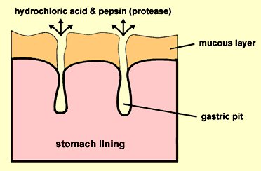
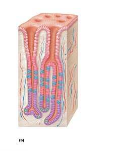
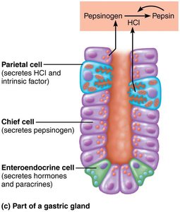
The stomach lining is composed entirely of goblet cells producing a protective mucus layer. Gastric pits lead to gastric glands, which contain several cell types:
Mucous neck cells: Secrete acidic mucin.
Parietal cells: Secrete HCl (activates pepsinogen, denatures proteins) and intrinsic factor (vitamin B12 absorption).
Chief cells: Secrete pepsinogen (converted to pepsin by HCl) and gastric lipase.
Enteroendocrine cells: Secrete hormones (gastrin, histamine, serotonin, somatostatin).
Stomach Protection Mechanisms
The stomach protects itself from its harsh acidic environment by:
Producing a thick, bicarbonate-rich mucus layer.
Having tight junctions between epithelial cells to prevent leakage of gastric juice.
Rapidly replacing surface epithelial cells (every 3–6 days) via stem cells at the gastric pit/gland junction.