 Back
BackDigestive System: Structure, Function, and Processes
Study Guide - Smart Notes
 Tailored notes based on your materials, expanded with key definitions, examples, and context.
Digestive System Overview
Introduction to the Digestive System
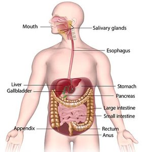
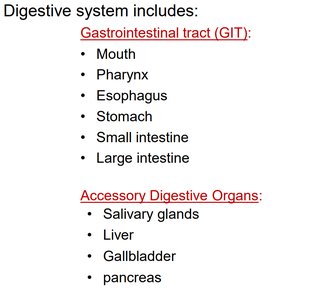
The digestive system is responsible for breaking down food into nutrients, which the body uses for energy, growth, and cell repair. It consists of the gastrointestinal tract (GIT) and accessory digestive organs. The GIT is a continuous tube running from the mouth to the anus, while accessory organs assist in digestion through secretion of enzymes and other substances.
Gastrointestinal tract (GIT): Mouth, Pharynx, Esophagus, Stomach, Small intestine, Large intestine
Accessory Digestive Organs: Salivary glands, Liver, Gallbladder, Pancreas
Major Food Groups and Their Digestion
Macromolecules in Food
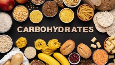
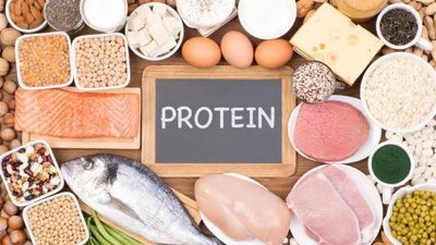
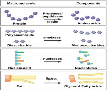
Food contains four major classes of macromolecules: carbohydrates, proteins, lipids (fats), and nucleic acids. Each is broken down by specific enzymes into absorbable monomers.
Carbohydrates: Broken down into monosaccharides (e.g., glucose)
Proteins: Broken down into amino acids
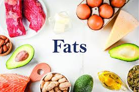
Fats (Lipids): Broken down into fatty acids and glycerol
Nucleic Acids: Broken down into nucleotides
Enzymatic Breakdown of Macromolecules
Enzymes catalyze the hydrolysis of macromolecules into their monomers:
Macromolecule | Enzyme | Product |
|---|---|---|
Protein | Proteases/pepsins | Amino acids |
Polysaccharide | Amylases | Monosaccharides |
Nucleic acid | Nucleases | Nucleotides |
Fat | Lipases | Glycerol, Fatty acids |
Processes of Digestion
Major Digestive Processes
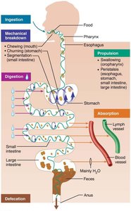
The digestive system carries out six essential processes:
Ingestion: Taking food into the mouth
Mechanical breakdown: Chewing (mouth), churning (stomach), segmentation (small intestine)
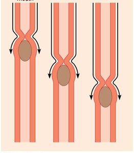
Propulsion: Swallowing and peristalsis move food through the GIT
Chemical digestion: Enzymatic breakdown of food
Absorption: Transport of digested nutrients into blood or lymph
Defecation: Elimination of indigestible substances as feces
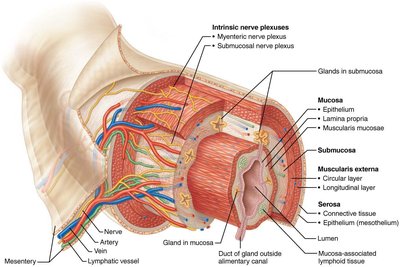
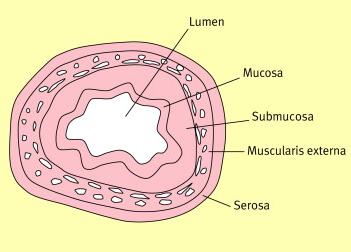
Histology of the Gastrointestinal Tract
General Structure of the GI Tract Wall
The wall of the GI tract consists of four main layers, each with specialized functions:
Mucosa: Innermost layer; contains epithelium, lamina propria, and muscularis mucosae. Functions in secretion, absorption, and protection.
Submucosa: Connective tissue with blood vessels, lymphatics, and nerves.
Muscularis externa: Smooth muscle (circular and longitudinal layers) responsible for peristalsis and segmentation. Skeletal muscle is present in the mouth, pharynx, upper esophagus, and anus for voluntary control.
Serosa: Outermost layer; visceral peritoneum covering the organs.
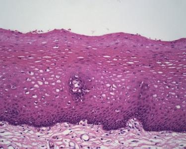
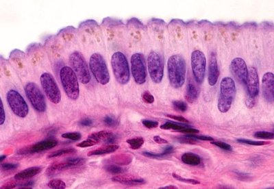
Types of Epithelium in the GI Tract
Stratified squamous epithelium: Found in the mouth, esophagus, and anal canal; protects against abrasion.
Simple columnar epithelium: Found in the stomach, small intestine, and large intestine; specialized for secretion and absorption.
Peristalsis
Peristalsis is the involuntary, rhythmic contraction of the muscularis externa that propels food through the digestive tract.
Mouth and Associated Structures
Oral Cavity (Mouth)
The mouth is the entry point for food and the site of both mechanical and chemical digestion. It contains the tongue, teeth, and is lined by a mucous membrane. The palate forms the roof, and the uvula helps prevent food from entering the nasal cavity during swallowing.
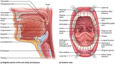
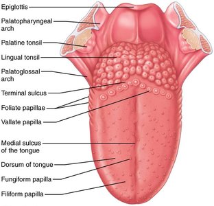
Tongue
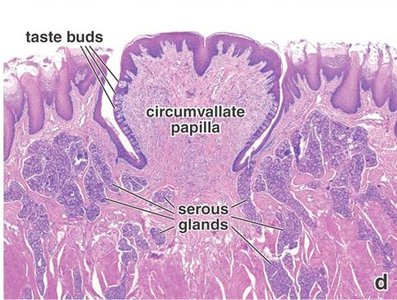
The tongue is a muscular organ that manipulates food, mixes it with saliva, and forms a bolus for swallowing. It contains taste buds for sensory detection and assists in speech articulation.
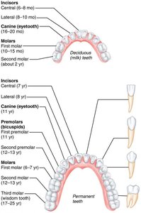
Teeth
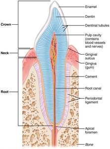
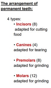
Teeth are essential for the mechanical breakdown of food. Each tooth has three regions: crown, neck, and root. The arrangement and types of teeth reflect their functions in cutting, tearing, and grinding food.
Incisors (8): Cutting food
Canines (4): Tearing food
Premolars (8): Grinding food
Molars (12): Grinding food
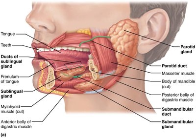
Salivary Glands
There are three pairs of major salivary glands: parotid, submandibular, and sublingual. They secrete saliva, which contains enzymes (salivary amylase, lingual lipase) and antimicrobial agents (lysozyme) that initiate chemical digestion and protect the oral cavity.
Summary of Digestion in the Mouth
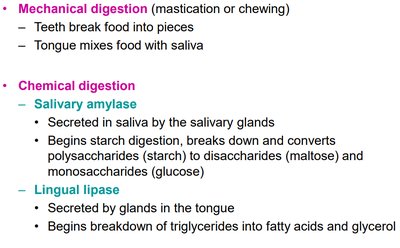
Mechanical digestion: Mastication (chewing) by teeth and mixing by tongue
Chemical digestion: Salivary amylase (starch to maltose), lingual lipase (triglycerides to fatty acids and glycerol)
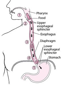
Pharynx and Esophagus
Pharynx
The pharynx is a muscular tube that serves as a passageway for both food (to the esophagus) and air (to the larynx). It connects the nasal and oral cavities to the esophagus and larynx.
Esophagus
The esophagus is a muscular tube that transports food from the pharynx to the stomach. It passes through the diaphragm and is regulated by the upper and lower esophageal sphincters. The upper third contains skeletal muscle, the middle third a mix of skeletal and smooth muscle, and the lower third smooth muscle only. No digestion occurs in the esophagus; its main function is propulsion.
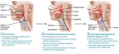
Swallowing (Deglutition)
Swallowing is a complex process involving voluntary and involuntary phases. The tongue pushes the bolus to the oropharynx (voluntary), followed by reflexive closure of the nasopharynx and larynx, and peristaltic movement of the bolus down the esophagus (involuntary).
Stomach
Structure and Function
The stomach is a J-shaped organ in the upper left abdomen. It stores, mixes, and digests food, converting it into chyme. The stomach has three regions (fundus, body, pylorus) and is bounded by the cardiac and pyloric sphincters. Its wall contains three muscle layers (circular, longitudinal, oblique) for churning food.
Gastric Glands and Secretions
The stomach mucosa contains gastric glands with specialized cells:
Mucous neck cells: Secrete mucus for lubrication
Parietal cells: Secrete hydrochloric acid (HCl) and intrinsic factor (for vitamin B12 absorption)
Chief cells: Secrete pepsinogen (converted to pepsin for protein digestion) and gastric lipase
G cells: Secrete gastrin hormone (stimulates gastric activity)
Functions of the Stomach
Liquefies food into chyme
Begins protein digestion (pepsin)
Begins lipid digestion (lingual and gastric lipase)
Continues starch digestion (salivary amylase)
Secretes intrinsic factor for vitamin B12 absorption
Absorbs some drugs and alcohol
Kills microbes with acidic gastric juice
Small Intestine
Structure and Regions
The small intestine is the primary site for digestion and absorption. It extends from the pyloric sphincter to the ileocecal valve and is divided into three regions: duodenum, jejunum, and ileum. The inner surface has circular folds, villi, and microvilli to increase surface area for absorption.
Histology and Specialized Cells
Absorptive cells: Have microvilli; secrete brush border enzymes
Goblet cells: Secrete mucus
Paneth cells: Secrete lysozyme (antimicrobial)
Enteroendocrine cells: Secrete hormones (GIP, secretin, CCK)
Hormonal Regulation
Gastric inhibitory peptide (GIP): Inhibits stomach, stimulates insulin secretion
Secretin: Inhibits stomach, stimulates bicarbonate secretion from pancreas and bile from liver
Cholecystokinin (CCK): Inhibits stomach, stimulates pancreatic enzyme secretion and bile release from gallbladder
Absorption in the Small Intestine
Monosaccharides, amino acids, and short-chain fatty acids are absorbed into blood capillaries
Long-chain fatty acids and fat-soluble vitamins (A, D, E, K) are absorbed into lacteals (lymphatic capillaries)
Vitamin B12 is absorbed with intrinsic factor
Large Intestine
Structure and Regions
The large intestine absorbs water and electrolytes, forming and storing feces. It consists of the cecum (with appendix), ascending colon, transverse colon, descending colon, sigmoid colon, rectum, and anal canal. The anal canal is guarded by internal (involuntary) and external (voluntary) sphincters.
Functions
Absorption of water and electrolytes
Formation and storage of feces
Host to beneficial bacteria (e.g., Bacteroides, Bifidobacterium, Escherichia coli)
Liver, Gallbladder, and Pancreas
Liver
The liver is the largest gland in the body, composed of hexagonal lobules with a central vein. Hepatocytes (liver cells) process nutrients, detoxify substances, and produce bile. Blood from the hepatic artery (oxygenated) and portal vein (nutrient-rich) mixes in sinusoids and drains into the central vein.
Bile and the Biliary System
Bile is a yellow-green fluid produced by hepatocytes, stored in the gallbladder, and released into the duodenum. It contains bile salts (for fat emulsification), cholesterol, phospholipids, and bile pigments (waste products). Bile does not contain enzymes but is essential for fat digestion and absorption.
Gallbladder
The gallbladder stores and concentrates bile between meals. When chyme enters the small intestine, CCK stimulates the gallbladder to contract and release bile into the duodenum.
Pancreas
The pancreas has both endocrine (insulin, glucagon) and exocrine (digestive enzymes) functions. Pancreatic juice contains bicarbonate (to neutralize gastric acid) and enzymes (amylase, proteases, lipase, nucleases) for digestion. Secretion is regulated by neural (vagus nerve) and hormonal (secretin, CCK) mechanisms.
Summary Table: Digestive Enzymes
Enzyme | Origin | Site of Action | Substrate | Product |
|---|---|---|---|---|
Salivary amylase | Salivary glands | Mouth | Starch | Maltose, glucose |
Pepsin | Stomach (chief cells) | Stomach | Proteins | Peptides |
Pancreatic amylase | Pancreas | Small intestine | Starch | Maltose, glucose |
Trypsin, chymotrypsin, carboxypeptidase | Pancreas | Small intestine | Proteins, peptides | Amino acids |
Pancreatic lipase | Pancreas | Small intestine | Triglycerides | Fatty acids, glycerol |
Nucleases | Pancreas | Small intestine | Nucleic acids | Nucleotides |
Brush border enzymes | Small intestine | Small intestine | Disaccharides, peptides | Monosaccharides, amino acids |
Key Concepts for Study
Know the structure and function of each region of the digestive tract, including specialized cells and secretions.
Understand the role of accessory organs (liver, gallbladder, pancreas) in digestion.
Be able to classify digestive enzymes by their origin, site of action, and substrate.
Recognize the four major macromolecules and their monomers.
