 Back
BackDigestive System: Structure, Function, and Processes
Study Guide - Smart Notes
 Tailored notes based on your materials, expanded with key definitions, examples, and context.
Digestive System Overview
Organization of the Digestive System
The digestive system is divided into two main groups: the alimentary canal (gastrointestinal tract) and accessory digestive organs. The alimentary canal is a continuous muscular tube that digests food and absorbs nutrients, while accessory organs assist in the digestive process through secretion and mechanical means.
Alimentary canal: Mouth, pharynx, esophagus, stomach, small intestine, large intestine
Accessory organs: Teeth, tongue, gallbladder, salivary glands, liver, pancreas
Digestive Processes
Six Essential Activities
Food processing in the digestive system involves six coordinated activities:
Ingestion: Taking food into the digestive tract
Propulsion: Moving food through the tract (includes swallowing and peristalsis)
Mechanical breakdown: Physically preparing food for digestion (chewing, churning, segmentation)
Digestion: Enzymatic breakdown of complex food molecules
Absorption: Passage of digested end products into blood or lymph
Defecation: Elimination of indigestible substances as feces
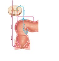
Regulation of Digestive Activity
GI Tract Regulatory Mechanisms
Digestive activity is regulated by neural and hormonal mechanisms. Mechanoreceptors and chemoreceptors in the GI tract respond to stretch, osmolarity, pH, and the presence of substrates, initiating reflexes that control glandular secretion and smooth muscle activity.
Short reflexes: Mediated by the enteric nervous system (gut brain)
Long reflexes: Involve the central nervous system and autonomic nerves
Hormonal control: Hormones from the stomach and small intestine regulate secretion and motility
Anatomy of the Digestive Tract
Peritoneum and Peritoneal Cavity
The peritoneum is a serous membrane lining the abdominal cavity and covering abdominal organs. The peritoneal cavity contains lubricating fluid, allowing organs to move smoothly. The mesentery is a double layer of peritoneum that supports organs and provides routes for blood vessels, lymphatics, and nerves.
Retroperitoneal organs: Located posterior to the peritoneum
Intraperitoneal organs: Surrounded by peritoneum
Histology of the Alimentary Canal
The alimentary canal wall consists of four basic layers (tunics):
Mucosa: Secretes mucus, digestive enzymes, and hormones; absorbs nutrients; protects against pathogens
Submucosa: Areolar connective tissue with blood vessels, lymphatics, and nerves
Muscularis externa: Responsible for segmentation and peristalsis; contains inner circular and outer longitudinal muscle layers
Serosa: Visceral peritoneum; replaced by adventitia in the esophagus
Enteric Nervous System
The enteric nervous system is the intrinsic nerve supply of the alimentary canal, containing more neurons than the spinal cord. It regulates GI tract motility and secretion, and is influenced by the autonomic nervous system.
Submucosal plexus: Controls glandular secretion and local blood flow
Myenteric plexus: Controls GI tract motility
Sympathetic stimulation: Inhibits digestive activity
Parasympathetic stimulation: Stimulates digestive activity
Functional Anatomy of the Upper GI Tract
Mouth, Lips, Cheeks, and Palate
The oral cavity is lined with stratified squamous epithelium and bounded by lips, cheeks, palate, and tongue. The hard palate provides a rigid surface for food manipulation, while the soft palate closes off the nasopharynx during swallowing.
Tongue
The tongue is a muscular organ that repositions and mixes food, forms the bolus, and initiates swallowing, speech, and taste. It contains various papillae (filiform, fungiform, circumvallate, foliate) with different functions, including taste sensation and friction.
Lingual lipase: Fat-digesting enzyme secreted by serous cells beneath papillae
Salivary Glands and Saliva
Salivary glands produce saliva, which cleanses the mouth, dissolves food chemicals, moistens food, and begins starch digestion. Saliva contains water, electrolytes, enzymes (amylase, lingual lipase), mucin, and antimicrobial agents.
Control of salivation: Parasympathetic stimulation increases secretion; sympathetic stimulation inhibits it
Teeth and Tooth Structure
Teeth are essential for mechanical digestion. Humans have two sets: primary (deciduous) and permanent teeth. Tooth structure includes the crown (covered by enamel), root (embedded in jawbone), dentin, pulp cavity, and periodontal ligament.
Pharynx and Esophagus
The pharynx and esophagus transport food from the mouth to the stomach. The esophagus is a muscular tube with specialized sphincters to prevent backflow and facilitate swallowing.
Digestive Processes in the Mouth and Stomach
Mouth
Mechanical digestion (mastication) and chemical digestion (salivary amylase and lingual lipase) begin in the mouth. Swallowing (deglutition) involves coordinated muscle actions in the buccal and pharyngeal-esophageal phases.
Stomach: Gross and Microscopic Anatomy
The stomach stores and digests food, converting it to chyme. It has specialized regions (cardia, fundus, body, pylorus) and is tethered by mesenteries (lesser and greater omentum). The stomach wall contains three muscle layers for churning and mixing food.
Mucosa: Contains gastric pits and glands that secrete gastric juice
Gastric glands: Mucous neck cells, parietal cells (HCl, intrinsic factor), chief cells (pepsinogen, lipase), enteroendocrine cells (hormones)
Gastric Secretion and Regulation
Gastric secretion is regulated by neural (vagus nerve) and hormonal (gastrin) mechanisms. There are three phases: cephalic (reflex), gastric, and intestinal. HCl formation involves active transport of H+ and Cl– ions, resulting in an alkaline tide in the blood.
Digestive Processes in the Stomach
Physical digestion: Churning and mixing of food
Chemical digestion: Pepsin digests proteins; lingual lipase acts on fats
Absorption: Limited to lipid-soluble substances (alcohol, aspirin)
Intrinsic factor: Essential for vitamin B12 absorption
Small Intestine: Structure and Function
Gross Anatomy
The small intestine is the major site of digestion and absorption, extending from the pyloric sphincter to the ileocecal valve. It is divided into the duodenum, jejunum, and ileum.
Structural Modifications for Absorption
Surface area is increased by circular folds, villi, and microvilli (brush border), enhancing nutrient absorption. Intestinal crypts contain secretory and immune cells.
Intestinal Juice
Intestinal juice is secreted in response to distension or irritation, facilitating nutrient absorption. It is slightly alkaline and enzyme-poor, with enzymes located in the brush border.
Accessory Digestive Organs
Liver and Gallbladder
The liver produces bile, which emulsifies fats. The gallbladder stores and concentrates bile, releasing it in response to cholecystokinin (CCK). Bile salts are recycled via enterohepatic circulation.
Liver lobules: Functional units containing hepatocytes, portal triads, and sinusoids with Kupffer cells
Bile composition: Bile salts, bilirubin, cholesterol, phospholipids, electrolytes
Pancreas
The pancreas has both endocrine (insulin, glucagon) and exocrine (pancreatic juice) functions. Pancreatic juice contains enzymes for digestion of all macronutrients and bicarbonate to neutralize acidic chyme.
Protease activation: Occurs in the duodenum (trypsinogen to trypsin, etc.)
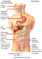
Digestion and Absorption
Chemical Digestion
Enzymatic hydrolysis breaks down macromolecules into absorbable monomers:
Carbohydrates: Digested by amylases and brush border enzymes to monosaccharides
Proteins: Digested by pepsin, pancreatic proteases, and brush border enzymes to amino acids
Lipids: Emulsified by bile salts, digested by pancreatic lipases to fatty acids and monoglycerides
Nucleic acids: Digested by pancreatic and brush border enzymes to nucleotide components
Absorption Mechanisms
Carbohydrates: Absorbed as monosaccharides via secondary active transport and facilitated diffusion
Proteins: Absorbed as amino acids, dipeptides, and tripeptides via active transport
Lipids: Absorbed as micelles, reassembled into chylomicrons, and transported via lacteals
Nucleic acids: Absorbed by active transport
Vitamins: Fat-soluble vitamins absorbed with lipids; water-soluble vitamins by diffusion or transporters; vitamin B12 with intrinsic factor
Electrolytes: Absorbed by active and passive mechanisms; iron and calcium absorption regulated by need
Water: Absorbed by osmosis, coupled with solute uptake
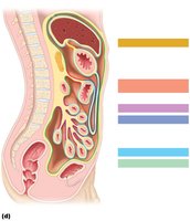
Large Intestine: Structure and Function
Gross Anatomy
The large intestine absorbs water, electrolytes, and vitamins produced by bacteria, and compacts indigestible residues into feces. It consists of the cecum, appendix, colon (ascending, transverse, descending, sigmoid), rectum, and anal canal.
Teniae coli: Longitudinal muscle bands
Haustra: Pocketlike sacs
Epiploic appendages: Fat-filled pouches
Bacterial Flora
Bacteria in the colon synthesize vitamins, ferment indigestible carbohydrates, and contribute to gas production. They also play a role in immune function.
Digestive Processes in the Large Intestine
Absorption: Water, electrolytes, and vitamins
Propulsion: Haustral contractions and mass movements move feces toward the rectum
Defecation: Initiated by stretch receptors, involving spinal reflexes and voluntary control
Aging and the Digestive System
Developmental and Age-Related Changes
With aging, digestive activity declines, absorption becomes less efficient, and peristalsis slows. Common issues include diverticulosis, fecal incontinence, and increased risk of GI cancers. Regular dental and medical exams are important for early detection and prevention.
Summary Table: Digestion and Absorption of Major Nutrients
Nutrient | Enzymes Involved | Absorption Mechanism |
|---|---|---|
Carbohydrates | Salivary amylase, pancreatic amylase, brush border enzymes | Secondary active transport (Na+), facilitated diffusion |
Proteins | Pepsin, pancreatic proteases, brush border enzymes | Active transport (Na+), H+-dependent cotransport |
Lipids | Bile salts, pancreatic lipase | Micelles, diffusion, chylomicrons via lacteals |
Nucleic acids | Pancreatic nucleases, brush border enzymes | Active transport |
