 Back
BackDigestive System: Structure, Function, and Processes
Study Guide - Smart Notes
 Tailored notes based on your materials, expanded with key definitions, examples, and context.
Digestive System Overview
Introduction
The digestive system is responsible for the intake, breakdown, absorption, and elimination of food. It consists of the alimentary canal and accessory organs, each playing a distinct role in digestion and nutrient absorption.
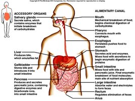
Alimentary Canal: The continuous tube through which food passes, including the mouth, pharynx, esophagus, stomach, small intestine, and large intestine.
Accessory Organs: Organs that aid digestion but do not have food pass through them directly, such as the teeth, tongue, salivary glands, liver, gallbladder, and pancreas.
Basic Divisions of the Digestive System
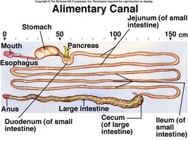
Alimentary Canal
The alimentary canal is the main passageway for food and waste. It is composed of several organs, each with specialized functions:
Mouth: Mechanical breakdown of food; begins chemical digestion of carbohydrates.
Pharynx: Moves food to the esophagus.
Esophagus: Transports food to the stomach.
Stomach: Secretes acid and enzymes; begins enzymatic digestion of proteins.
Small Intestine: Completes digestion; absorbs nutrients and electrolytes.
Large Intestine: Absorbs water; forms and expels feces.
Rectum and Anus: Regulates elimination of feces.
Accessory Organs
Salivary Glands: Secrete saliva containing enzymes for carbohydrate breakdown.
Liver: Produces bile for fat emulsification.
Gallbladder: Stores and concentrates bile.
Pancreas: Secretes digestive enzymes and bicarbonate into the small intestine.
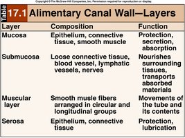
Layers of the Gastrointestinal (GI) Tract
Overview of GI Tract Wall
The GI tract wall is composed of four main layers, each with distinct functions:
Mucosa: Epithelium, connective tissue, and smooth muscle; functions in protection, secretion, and absorption.
Submucosa: Loose connective tissue, blood vessels, lymphatic vessels, and nerves; nourishes tissues and transports absorbed materials.
Muscularis (Muscular Layer): Smooth muscle fibers arranged in circular and longitudinal groups; responsible for movements such as peristalsis and segmentation.
Serosa: Epithelium and connective tissue; provides protection and lubrication.
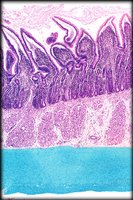
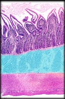
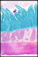
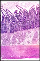
Table: Layers of the Alimentary Canal Wall
Layer | Composition | Function |
|---|---|---|
Mucosa | Epithelium, connective tissue, smooth muscle | Protection, secretion, absorption |
Submucosa | Loose connective tissue, blood vessels, lymphatic vessels, nerves | Nourishes surrounding tissues, transports absorbed materials |
Muscular layer | Smooth muscle fibers arranged in circular and longitudinal groups | Movements of the tube and its contents |
Serosa | Epithelium, connective tissue | Protection, lubrication |
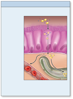
Mucosa Layer
Structure and Function
The mucosa is the innermost lining of the GI tract, responsible for secretion, protection, and absorption. It consists of three sublayers:
Epithelium: Absorbs nutrients and secretes mucus and enzymes.
Lamina propria: Connective tissue supporting the epithelium.
Muscularis mucosae: Thin layer of smooth muscle aiding local movements.
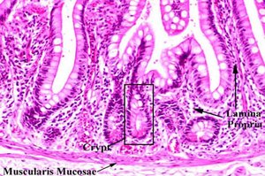
Submucosa Layer
Structure and Function
The submucosa is a dense connective tissue layer containing blood vessels, lymphatic vessels, lymph nodes, and nerves. It supports the mucosa and facilitates transport of absorbed nutrients.
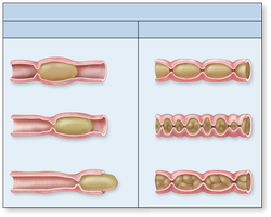
Muscularis Externa
Structure and Function
The muscularis externa typically consists of two layers of smooth muscle: an inner circular layer and an outer longitudinal layer. These layers are responsible for:
Sphincters: Control passage of materials.
Peristalsis: Wave-like contractions moving food along the GI tract.
Segmentation: Mixing movements that increase contact with digestive enzymes.
Serosa and Adventitia
Structure and Function
The serosa is the outermost layer for intraperitoneal organs, also known as the visceral peritoneum. Organs outside the peritoneal cavity have an adventitia, composed of areolar connective tissue. Both provide protection and lubrication.
General Functions of the Digestive System
Six Main Functions
Ingestion: Introduction of food and liquids into the oral cavity.
Motility: Muscular contractions for mixing and moving materials.
Secretion: Production and release of digestive fluids.
Digestion: Breakdown of food (mechanical and chemical).
Absorption: Transport of nutrients into blood or lymph.
Elimination: Expulsion of indigestible components.
Regulation of Digestive System Processes
Receptors and Nervous Control
Receptors in the GI tract monitor changes and relay sensory input to the central nervous system. Autonomic motor output coordinates secretory and muscular activity (long reflexes), while local reflexes (short reflexes) are managed by the enteric nervous system.
Hormonal Control
Gastrin: From the stomach, stimulates gastric activity.
Secretin: From the small intestine, stimulates bicarbonate secretion.
Cholecystokinin (CCK): From the small intestine, stimulates bile and pancreatic juice release.
Summary Table: Digestive System Functions
Process | Location | Purpose |
|---|---|---|
Ingestion | Mouth | Intake of food |
Propulsion | Pharynx, esophagus | Movement of food |
Mechanical Digestion | Mouth, stomach, small intestine | Physical breakdown |
Chemical Digestion | Stomach, small intestine | Enzymatic breakdown |
Absorption | Small intestine, large intestine | Uptake of nutrients |
Defecation | Rectum, anus | Elimination of waste |
Conclusion
The digestive system is a complex network of organs and tissues that work together to ensure the proper breakdown, absorption, and elimination of food. Understanding its structure and function is essential for comprehending human physiology and maintaining health.
