 Back
BackDigestive System: Structure, Function, and Regulation
Study Guide - Smart Notes
 Tailored notes based on your materials, expanded with key definitions, examples, and context.
The Digestive System
Overview and Divisions
The digestive system is responsible for acquiring nutrients from the environment, which are used for anabolism (synthesis of essential compounds) and catabolism (energy production). It consists of two main divisions: the digestive tract (alimentary canal) and accessory digestive organs.
Digestive tract: A muscular tube including the oral cavity, pharynx, esophagus, stomach, small intestine, large intestine, and anus.
Accessory organs: Teeth, tongue, salivary glands, liver, gallbladder, and pancreas.
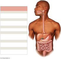
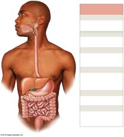
Major Processes of the Digestive System
Ingestion: Intake of food into the digestive tract.
Mechanical digestion and propulsion: Physical breakdown and movement of food.
Chemical digestion: Enzymatic breakdown of food into absorbable molecules.
Secretion: Release of water, acids, enzymes, buffers, and salts.
Absorption: Movement of nutrients, water, and electrolytes into the blood or lymph.
Defecation: Elimination of indigestible substances as feces.
Anatomy of the Digestive Tract
Mesenteries
Mesenteries are double sheets of peritoneal membrane that support and stabilize the digestive organs, providing a route for blood vessels, nerves, and lymphatics.
Lesser omentum: Stabilizes the position of the stomach and provides a route for blood vessels entering the liver.
Greater omentum: Protects the abdominal viscera and stores fat.
Mesocolon: Attaches parts of the large intestine to the posterior abdominal wall.
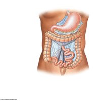
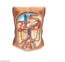
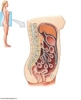
Histology of the Digestive Tract
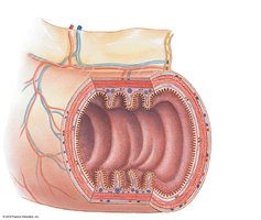
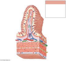
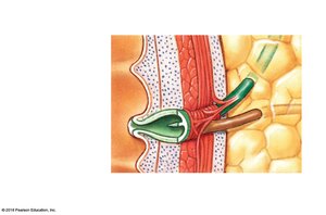
The wall of the digestive tract consists of four major layers:
Mucosa: Inner lining; mucous membrane with epithelium, lamina propria, and muscularis mucosae.
Submucosa: Dense irregular connective tissue with blood vessels, lymphatics, and sometimes glands.
Muscular layer: Smooth muscle (inner circular and outer longitudinal layers) responsible for peristalsis and segmentation.
Serosa: Serous membrane covering most of the digestive tract; replaced by adventitia in some regions.
Motility: Peristalsis and Segmentation
Movement of materials is achieved by coordinated contractions of smooth muscle:
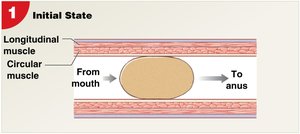
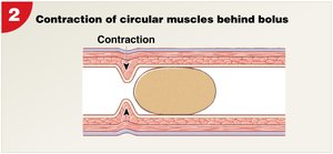
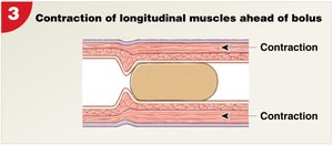
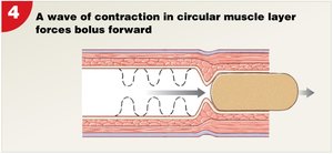
Peristalsis: Waves of muscular contractions that move a bolus along the tract.
Segmentation: Cycles of contraction that mix and churn contents without net movement.
Regulation of Digestive Functions
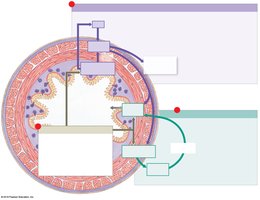
Local factors: Changes in pH, physical distortion, or chemical signals in the tract wall.
Neural mechanisms: Short (myenteric) and long reflexes coordinate motility and secretion.
Hormonal mechanisms: Enteroendocrine cells release hormones that regulate digestion.
The Oral Cavity
Functions and Anatomy
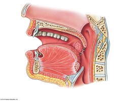
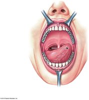
The oral cavity is involved in sensory analysis, mechanical digestion, lubrication, and limited chemical digestion of carbohydrates and lipids.
Teeth: Responsible for mastication (chewing).
Tongue: Assists in mechanical digestion, manipulation, sensory analysis, and secretion of mucins and lingual lipase.
Salivary glands: Produce saliva for lubrication, digestion, and protection.
Teeth and Dentition
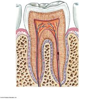
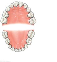
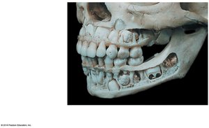
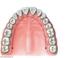
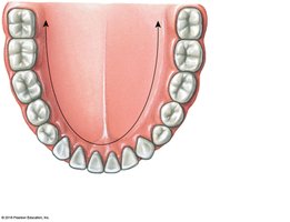
Teeth are specialized for cutting, tearing, and grinding food. Humans have two sets of teeth: deciduous (primary) and permanent.
Incisors: Cutting
Canines (cuspids): Tearing
Premolars (bicuspids) and molars: Grinding
Salivary Glands and Saliva
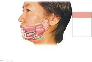
Three major pairs of salivary glands secrete saliva into the oral cavity:
Parotid glands: Produce serous secretion with salivary amylase.
Sublingual glands: Produce mucus for lubrication.
Submandibular glands: Secrete buffers, mucins, and amylase.
The Pharynx and Esophagus
Pharynx
The pharynx is a common passageway for food, liquids, and air, divided into nasopharynx, oropharynx, and laryngopharynx.
Esophagus
The esophagus is a muscular tube (about 25 cm long) that conveys food and liquids to the stomach. It is lined by mucosa, submucosa, and muscular layers, and is anchored by adventitia.
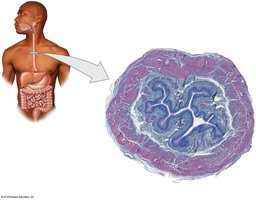
Deglutition (Swallowing)
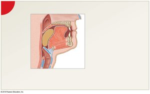
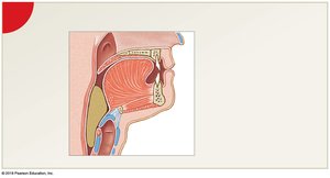
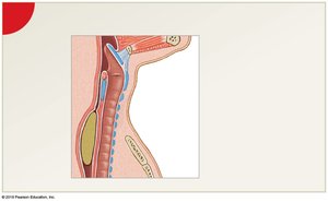
Swallowing involves three phases:
Buccal phase: Voluntary; bolus is compressed against the palate and moved into the oropharynx.
Pharyngeal phase: Involuntary; bolus passes through the pharynx as the epiglottis closes the glottis.
Esophageal phase: Involuntary; peristalsis moves the bolus to the stomach.
The Stomach
Structure and Function
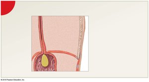
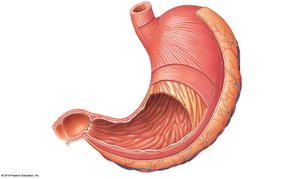
The stomach temporarily stores ingested food, performs mechanical digestion, and initiates chemical digestion of proteins and lipids. The partially digested mixture is called chyme.
Regions: Cardia, fundus, body, pyloric part
Rugae: Folds in the mucosa that allow expansion
Muscularis externa: Includes an additional oblique muscle layer
Gastric Glands and Secretions
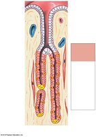
Gastric glands contain several cell types:
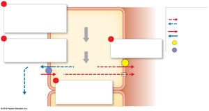
Parietal cells: Secrete intrinsic factor and hydrochloric acid (HCl)
Chief cells: Secrete pepsinogen (converted to pepsin by HCl)
Enteroendocrine cells: Secrete hormones such as gastrin and somatostatin
Regulation of Gastric Activity
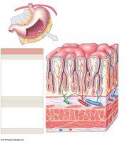
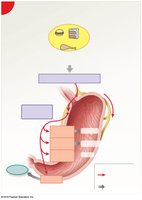
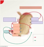
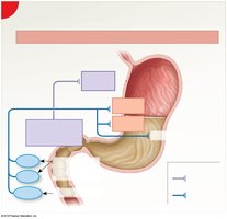
Gastric activity is regulated in three overlapping phases:
Cephalic phase: Initiated by sight, smell, taste, or thought of food; mediated by the CNS.
Gastric phase: Begins with arrival of food in the stomach; involves neural, hormonal, and local responses.
Intestinal phase: Begins when chyme enters the small intestine; inhibits gastric activity.
Accessory Digestive Organs
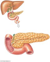
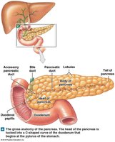
Pancreas
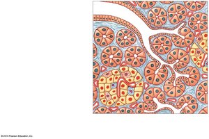
The pancreas has both endocrine and exocrine functions:
Endocrine cells: Secrete insulin and glucagon.
Exocrine cells: Secrete pancreatic juice containing digestive enzymes and bicarbonate.
Liver
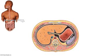
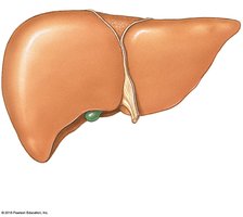
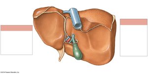
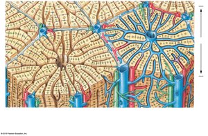
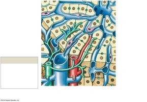
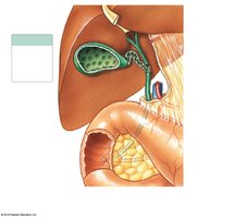
The liver is the largest visceral organ and performs metabolic, hematological, and synthetic functions. It is divided into lobes and receives blood from the hepatic artery and portal vein.
Hepatocytes: Adjust nutrient levels, detoxify blood, and produce bile.
Bile: Essential for emulsification of lipids in the small intestine.
Gallbladder
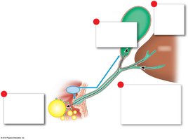
The gallbladder stores and concentrates bile, releasing it into the duodenum in response to cholecystokinin (CCK).
The Small Intestine
Structure and Function
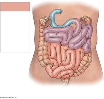
The small intestine is the primary site for chemical digestion and absorption of nutrients. It consists of three segments: duodenum, jejunum, and ileum.
Duodenum: Receives chyme and digestive secretions.
Jejunum: Site of most digestion and absorption.
Ileum: Ends at the ileocecal valve, controlling entry into the large intestine.
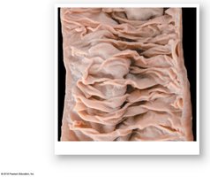
Histology and Absorption
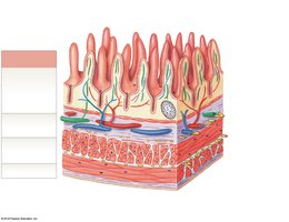
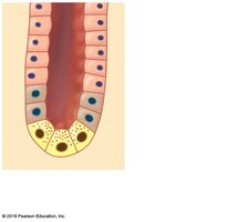
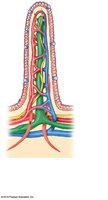
The small intestine has specialized structures to increase surface area for absorption:
Circular folds (plicae circulares)
Villi: Fingerlike projections of mucosa
Microvilli: Form the brush border on epithelial cells
The Large Intestine
Structure and Function
The large intestine absorbs water, electrolytes, and vitamins produced by bacteria, compacts feces, and stores fecal material prior to defecation. It consists of the cecum, colon (ascending, transverse, descending, sigmoid), rectum, and anus.
Haustra: Pouches that permit expansion
Teniae coli: Longitudinal muscle bands
Omental appendices: Fat-filled sacs
Functions and Microbiome
Absorption of water, bile salts, vitamins, and organic wastes
Compaction and storage of feces
Microbiome produces vitamins (K, biotin, B5) and gases (flatus)
Motility and Defecation Reflex
Movement through the large intestine is slow, allowing water absorption. Mass movements and reflexes (gastroileal, gastroenteric, defecation reflex) coordinate elimination.
Chemical Digestion and Absorption
Overview of Nutrient Digestion
Carbohydrates: Broken down by amylases and brush border enzymes to monosaccharides
Lipids: Emulsified by bile salts, digested by lipases, absorbed as fatty acids and monoglycerides
Proteins: Digested by pepsin, pancreatic proteases, and brush border enzymes to amino acids
Water and Electrolyte Absorption
Most water is absorbed in the small intestine, with additional reabsorption in the colon. Electrolytes and vitamins are absorbed by specific transport mechanisms.
Summary Table: Digestive Tract Layers
Layer | Main Features |
|---|---|
Mucosa | Epithelium, lamina propria, muscularis mucosae; secretion and absorption |
Submucosa | Connective tissue, blood/lymph vessels, glands, submucosal plexus |
Muscular layer | Smooth muscle (circular and longitudinal), myenteric plexus; motility |
Serosa/Adventitia | Serous membrane (serosa) or connective tissue (adventitia) |
Additional info: This guide covers the main anatomical and physiological features of the digestive system, including histology, motility, regulation, and the roles of accessory organs. For more detail on enzymatic pathways and absorption mechanisms, refer to advanced physiology texts.
