 Back
BackDigestive System: Structure, Function, and Regulation 26-1
Study Guide - Smart Notes
 Tailored notes based on your materials, expanded with key definitions, examples, and context.
Digestive System Overview
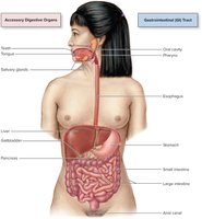
Main Functions of the Digestive System
The digestive system is responsible for processing food to extract and absorb nutrients, and for eliminating indigestible remains. Its main functions include:
Ingestion: Taking in food through the mouth.
Mechanical Breakdown: Physically breaking food into smaller pieces (chewing, churning, segmentation).
Chemical Digestion: Enzymatic breakdown of complex molecules into absorbable units.
Absorption: Transport of digested nutrients into blood or lymph.
Propulsion: Moving food through the alimentary canal (swallowing, peristalsis).
Defecation: Elimination of indigestible substances as feces.
Digestive Processes
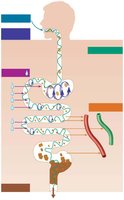
Six Essential Activities
Food processing in the digestive system involves six coordinated activities:
Ingestion: Entry of food into the digestive tract.
Propulsion: Movement of food, primarily by peristalsis (waves of muscle contraction and relaxation).
Mechanical Breakdown: Chewing, mixing, and segmentation to increase surface area for enzymes.
Chemical Digestion: Enzymatic breakdown of food into basic building blocks.
Absorption: Passage of nutrients from the GI tract into blood or lymph.
Defecation: Removal of indigestible substances.
Motility in the GI Tract
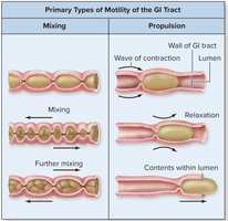
Motility refers to the movements that mix and propel contents through the digestive tract:
Mixing: Segmentation contractions mix food with digestive juices.
Propulsion: Peristalsis moves food along the tract.
Organization of the Digestive System
Peritoneum and Mesenteries
The digestive organs are located in the abdominopelvic cavity and are covered by serous membranes called the peritoneum:
Visceral Peritoneum: Covers external surfaces of most digestive organs.
Parietal Peritoneum: Lines the body wall.
Peritoneal Cavity: Fluid-filled space between the two peritoneal layers, reducing friction.
Mesentery: Double layer of peritoneum that supports organs, provides routes for vessels and nerves, and stores fat.
Retroperitoneal Organs: Located behind the peritoneum (e.g., most of the pancreas, duodenum, parts of the large intestine).
Histological Structure of the GI Tract
General Layers (Tunics) of the GI Tract
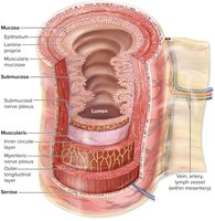
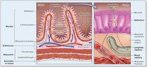
The walls of the alimentary canal (GI tract) are composed of four basic layers, or tunics:
Mucosa: Innermost layer; functions in secretion, absorption, and protection.
Epithelium: Mostly simple columnar cells for absorption and secretion.
Lamina Propria: Loose areolar connective tissue with blood vessels and lymphatics.
Muscularis Mucosae: Thin layer of smooth muscle.
Submucosa: Areolar connective tissue containing blood and lymphatic vessels, lymphoid follicles, and the submucosal nerve plexus.
Muscularis Externa: Responsible for segmentation and peristalsis; consists of inner circular and outer longitudinal muscle layers. Contains the myenteric nerve plexus.
Serosa: Outermost layer; visceral peritoneum formed from areolar connective tissue covered with mesothelium. In the esophagus, this is replaced by adventitia (dense connective tissue).
Absorption Across the Epithelial Lining
Absorption occurs primarily in the small intestine, where digested nutrients pass through the epithelial lining into blood and lymphatic vessels located in the lamina propria.
Control of Digestive System
Enteric Nervous System
The digestive system is regulated by its own intrinsic nervous system, the enteric nervous system (ENS), which consists of two main plexuses:
Submucosal Nerve Plexus: Regulates glands and smooth muscle in the mucosa.
Myenteric Nerve Plexus: Controls GI tract motility (movement).
Neural Reflex Pathways
Digestive activity is regulated by both local (intrinsic) and central (extrinsic) neural mechanisms:
Internal Stimuli: Changes in stretch, pH, nutrient, or solute concentration in the GI tract activate chemoreceptors, osmoreceptors, or mechanoreceptors.
Local Reflexes: Initiated by the ENS, affecting smooth muscle and glandular activity.
External Stimuli: Sight, smell, taste, or thought of food can trigger central nervous system responses that influence digestive activity.
Effectors: Smooth muscle and glands respond by altering contractile or secretory activity.
Summary Table: Layers of the GI Tract
Layer | Main Components | Functions |
|---|---|---|
Mucosa | Epithelium, lamina propria, muscularis mucosae | Secretion, absorption, protection |
Submucosa | Areolar connective tissue, blood/lymph vessels, submucosal plexus | Support, vascular supply, nerve supply |
Muscularis externa | Inner circular and outer longitudinal muscle, myenteric plexus | Motility (mixing, peristalsis) |
Serosa | Areolar connective tissue, mesothelium | Protection, reduces friction |
Key Terms and Concepts
Peristalsis: Coordinated, rhythmic contractions that propel food through the GI tract.
Segmentation: Localized contractions that mix food with digestive juices.
Retroperitoneal: Organs located behind the peritoneum.
Mesentery: Double layer of peritoneum supporting abdominal organs.
Enteric Nervous System: Intrinsic nerve network regulating digestive function.
Additional info: The digestive system is closely integrated with the circulatory and lymphatic systems for nutrient absorption and immune defense.
