 Back
Digestive System: Structure, Function, and Regulation
Study Guide - Smart Notes
 Tailored notes based on your materials, expanded with key definitions, examples, and context.
Organization of the Digestive System
Overview of Digestive System Structure
The digestive system is composed of the gastrointestinal (GI) tract and accessory digestive organs. The GI tract forms a continuous tube through which food passes and is processed, while accessory organs assist in digestion through mechanical and chemical means.
GI Tract Organs: Oral cavity, pharynx, esophagus, stomach, small intestine, large intestine, anus
Accessory Organs: Teeth, tongue, salivary glands, liver, gallbladder, pancreas
Function: Breakdown of food, absorption of nutrients, and elimination of waste
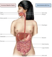
General Functions of the Digestive System
Six Main Functions
Ingestion: Intake of food and liquids into the oral cavity
Motility: Muscular contractions (voluntary and involuntary) that mix and move contents through the GI tract
Secretion: Release of digestive fluids (enzymes, acid, bile) to facilitate digestion
Digestion: Breakdown of food into smaller components
Mechanical digestion: Physical breakdown (chewing, mixing)
Chemical digestion: Enzymatic breakdown of macromolecules
Absorption: Movement of digested molecules, electrolytes, vitamins, and water from the GI tract into blood or lymph
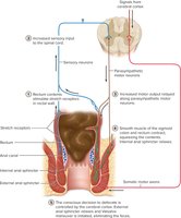
Elimination: Expulsion of indigestible substances as feces
Histological Organization of the GI Tract Wall
Four Concentric Layers (Tunics)
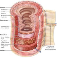
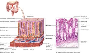
The GI tract wall consists of four main layers, each with specialized functions:
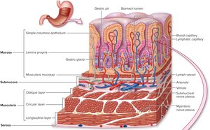
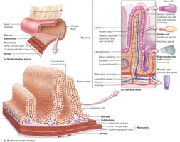
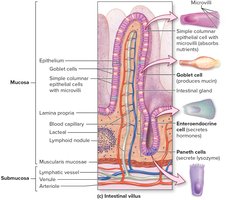
Mucosa: Innermost layer; includes epithelium (for secretion and absorption), lamina propria (areolar tissue, blood vessels, nerves), and muscularis mucosae (smooth muscle)
Submucosa: Areolar and dense irregular connective tissue; contains blood vessels, lymphatics, nerves (submucosal plexus), and MALT (gut-associated lymphoid tissue)
Muscularis: Two layers of smooth muscle (inner circular and outer longitudinal); responsible for peristalsis and mixing; contains myenteric nerve plexus
Adventitia/Serosa: Outermost layer; adventitia (areolar CT) outside peritoneal cavity, serosa (with visceral peritoneum) for intraperitoneal organs
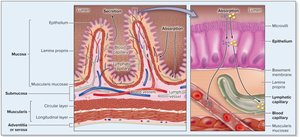
Absorption Across the Epithelial Lining
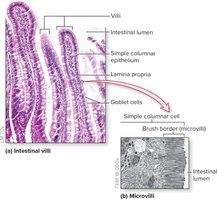
Absorption occurs primarily across the simple columnar epithelium of the mucosa, with nutrients entering blood and lymphatic capillaries in the lamina propria.
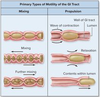
Motility: Mixing and Propulsion
Motility in the GI tract includes two primary types:
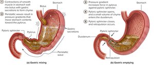
Mixing: Back-and-forth motion that blends ingested materials with secretions, increasing contact with digestive enzymes
Propulsion: Directional movement of contents through the GI tract, primarily by peristalsis (sequential muscular contractions)
Serous Membranes of the Abdominal Cavity
Peritoneum and Omenta
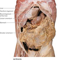
The peritoneum is a serous membrane lining the abdominopelvic cavity and covering abdominal organs. It consists of the parietal peritoneum (lining the abdominal wall) and the visceral peritoneum (covering organs). The peritoneal cavity is the potential space between these layers, containing lubricating serous fluid.
Omenta: Folds of peritoneum (greater and lesser omentum) that support and protect abdominal organs
Oral Cavity and Salivary Glands
Anatomy of the Oral Cavity
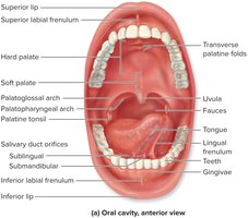
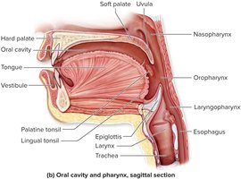
The oral cavity is the entrance to the GI tract and is divided into the vestibule (between gums, lips, cheeks) and the oral cavity proper (central to the teeth, leading to the oropharynx). It is lined by stratified squamous epithelium for protection against abrasion.
Salivary Glands
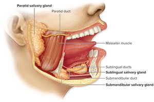
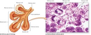
Salivary glands produce saliva, which moistens food, initiates digestion, and protects oral structures. There are intrinsic (within oral cavity) and extrinsic (parotid, submandibular, sublingual) glands. Saliva contains salivary amylase (starch digestion), lingual lipase (fat digestion), mucin, lysozyme, and IgA antibodies.
Pharynx and Esophagus
Gross Anatomy and Histology
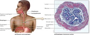
The pharynx is a muscular passageway for air and food, lined by nonkeratinized stratified squamous epithelium. The esophagus is a collapsible tube connecting the pharynx to the stomach, with superior and inferior esophageal sphincters regulating passage and preventing reflux.
Swallowing (Deglutition)
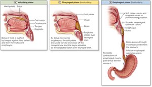
Swallowing involves three phases:
Voluntary phase: Bolus formed and pushed toward oropharynx
Pharyngeal phase: Involuntary; soft palate and uvula elevate, larynx rises, epiglottis covers airway
Esophageal phase: Involuntary; peristaltic waves move bolus to stomach
Stomach
Gross Anatomy and Regions
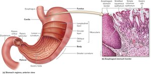
The stomach is a J-shaped organ with four regions: cardia, fundus, body, and pylorus. It serves as a reservoir for food, mixes contents to form chyme, and initiates protein and fat digestion. The pyloric sphincter regulates chyme entry into the duodenum.
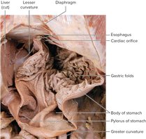
Histology of the Stomach
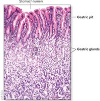
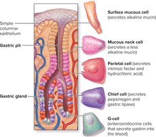
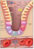
The stomach wall has three layers of smooth muscle (oblique, circular, longitudinal) for churning. The mucosa contains gastric pits leading to gastric glands, which house various secretory cells:
Surface mucous cells: Secrete alkaline mucin
Mucous neck cells: Secrete less alkaline mucin
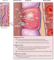
Parietal cells: Secrete intrinsic factor and hydrochloric acid (HCl)
Chief cells: Secrete pepsinogen and gastric lipase
G-cells: Secrete gastrin (hormone)
Formation of HCl
Parietal cells secrete HCl, which lowers stomach pH, denatures proteins, activates pepsinogen, and kills microbes.
Gastric Secretions and Regulation
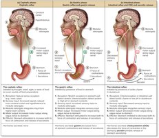
Gastric secretions are regulated in three phases:
Cephalic phase: Initiated by thought, smell, or sight of food; increases gastric activity
Gastric phase: Triggered by food in stomach; gastrin release enhances secretion and motility
Intestinal phase: Begins when chyme enters duodenum; CCK and secretin inhibit gastric activity
Lower GI Tract and Accessory Organs
Small and Large Intestine
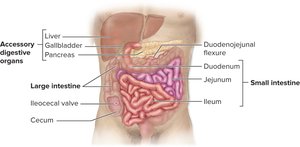
The small intestine (duodenum, jejunum, ileum) is the primary site for digestion and absorption. The large intestine absorbs water and electrolytes, forming and storing feces.
Small Intestine Structure
The small intestine has three main features that increase surface area for absorption:
Circular folds: Folds of mucosa and submucosa
Villi: Fingerlike projections of mucosa
Microvilli: Extensions of epithelial cell membranes (brush border)
Accessory Digestive Organs and Ducts
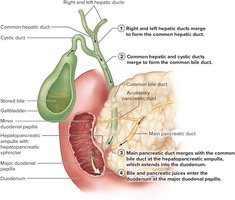
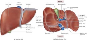
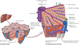
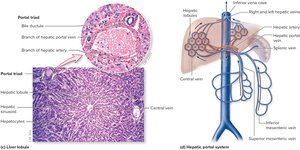
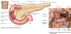
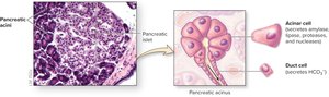
The liver produces bile, the gallbladder stores and concentrates bile, and the pancreas produces digestive enzymes and bicarbonate. These secretions enter the duodenum via a network of ducts (biliary apparatus and pancreatic ducts).
Large Intestine
Gross Anatomy and Function
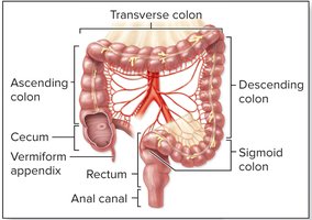
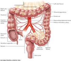
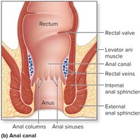
The large intestine consists of the cecum, colon (ascending, transverse, descending, sigmoid), rectum, and anal canal. It absorbs water and electrolytes, compacts feces, and houses gut microbiota.
Summary Table: Digestive Tract Wall Layers
Layer | Main Components | Function |
|---|---|---|
Mucosa | Epithelium, lamina propria, muscularis mucosae | Secretion, absorption, protection |
Submucosa | Connective tissue, blood/lymph vessels, nerves, MALT | Support, immune defense, innervation |
Muscularis | Inner circular and outer longitudinal smooth muscle | Motility (mixing, peristalsis) |
Adventitia/Serosa | Areolar connective tissue (serosa also has visceral peritoneum) | Protection, structural support |
Additional info: The notes above integrate and expand upon the provided material, ensuring a comprehensive, self-contained study guide for ANP college students. Images are included only where they directly reinforce the adjacent content.
