 Back
BackECG and Peripheral Circulation: Study Guide for ANP College Students
Study Guide - Smart Notes
 Tailored notes based on your materials, expanded with key definitions, examples, and context.
Cardiovascular System: Heart Sounds and Pulse Points
Heart Sounds and Auscultation
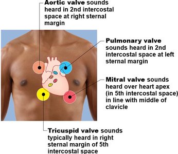
The four main heart sounds are auscultated at specific regions on the chest, corresponding to the locations of the heart valves. Understanding these locations is essential for clinical assessment of cardiac function.
Aortic valve: Heard in the 2nd intercostal space at the right sternal margin.
Pulmonary valve: Heard in the 2nd intercostal space at the left sternal margin.
Tricuspid valve: Typically heard in the right sternal margin of the 5th intercostal space.
Mitral valve: Heard over the heart apex (5th intercostal space, in line with the middle of the clavicle).
Pulse Points of the Arm
Pulse points are locations where arteries are close to the skin and can be palpated to assess heart rate and rhythm. The correct technique involves using the index and middle fingers, avoiding the thumb due to its own pulse.
Radial pulse: Thumb side of the wrist.
Brachial pulse: Inside of the elbow.
Ulnar pulse: Pinky side of the wrist.
Arteries: Pulses are felt in arteries, not veins, as arteries carry blood away from the heart and have strong pressure.
Peripheral Circulation: Major Arteries and Veins
Arteries of the Arm
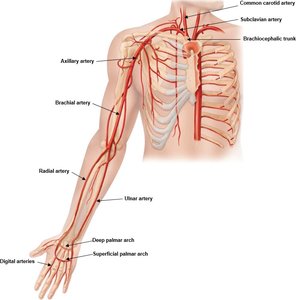
The arteries of the arm supply oxygenated blood to the upper limb. Knowledge of their locations is important for clinical procedures and pulse assessment.
Subclavian artery
Axillary artery
Brachial artery
Radial artery
Ulnar artery
Palmar arches (deep and superficial)
Digital arteries
Veins of the Arm
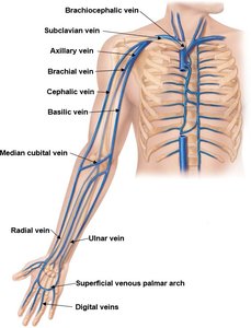
The veins of the arm return deoxygenated blood to the heart. They are important for venipuncture and intravenous access.
Brachiocephalic vein
Subclavian vein
Axillary vein
Brachial vein
Cephalic vein
Basilic vein
Median cubital vein
Radial vein
Ulnar vein
Superficial venous palmar arch
Digital veins
Arteries of the Leg
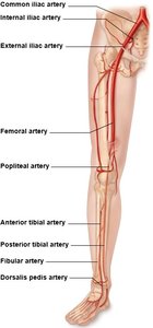
The arteries of the leg deliver oxygenated blood to the lower limb. Their identification is crucial for assessing peripheral circulation.
Common iliac artery
Internal and external iliac arteries
Femoral artery
Popliteal artery
Anterior and posterior tibial arteries
Fibular artery
Dorsalis pedis artery
Veins of the Leg
The veins of the leg are responsible for returning blood to the heart. They are commonly used for venous access and assessment of venous return.
Common iliac vein
Internal and external iliac veins
Femoral vein
Great saphenous vein (superficial)
Popliteal vein
Small saphenous vein
Fibular vein
Anterior tibial vein
Dorsalis pedis vein

Great Vessels of the Heart
Major Arteries and Branches
The great vessels are large arteries and veins that transport blood to and from the heart. Their identification is fundamental in cardiovascular anatomy.
Aorta: Main artery leaving the heart.
Brachiocephalic trunk: Branches into the right subclavian and right carotid arteries.
Left common carotid artery
Left subclavian artery
Pulmonary trunk: Branches into pulmonary arteries, carrying deoxygenated blood to the lungs.
Cardiac Conduction System
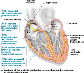
Anatomy of the Intrinsic Conduction System
The cardiac conduction system coordinates the heart's electrical activity, ensuring proper timing of contractions. The sequence of excitation is crucial for normal heart function.
Sinoatrial (SA) node: Pacemaker, generates impulses.
Atrioventricular (AV) node: Delays impulse, allows atria to contract.
AV bundle (Bundle of His): Connects atria to ventricles.
Bundle branches: Conduct impulses through interventricular septum.
Subendocardial conducting network (Purkinje fibers): Stimulates contractile cells of ventricles.
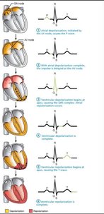
ECG Waves and Physiological Events
An electrocardiogram (ECG) records the electrical activity of the heart. Each wave corresponds to a specific physiological event.
P wave: Atrial depolarization.
QRS complex: Ventricular depolarization (and atrial repolarization).
T wave: Ventricular repolarization.
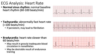
ECG Analysis: Heart Rate and Arrhythmias
Normal Sinus Rhythm and Arrhythmias
ECG analysis is used to assess heart rhythm and detect arrhythmias. Recognizing normal and abnormal patterns is essential for diagnosis.
Normal sinus rhythm: 60-100 beats/min, regular rhythm.
Tachycardia: >100 beats/min, abnormally fast.
Bradycardia: <60 beats/min, abnormally slow.
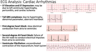
ST elevation/ST depression: May indicate ischemia or hypertrophy.
Tall QRS complexes: May indicate abnormal pacemaker or hypertrophy.
First-degree heart block: Slow conduction from atria to ventricles.
Second-degree AV block: Failure of AV node to conduct impulses.
Ventricular fibrillation: Uncoordinated contraction, heart quiver.
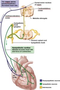
Extrinsic Control of Heart Rate
Autonomic Nervous System Regulation
The heart rate and force of contraction are regulated by the autonomic nervous system (ANS) via cardiac centers in the medulla oblongata.
Cardioacceleratory center (sympathetic): Increases heart rate and force via sympathetic trunk; stimulates SA node, AV node, heart muscle, and coronary arteries.
Cardioinhibitory center (parasympathetic): Decreases heart rate via the vagus nerve; inhibits SA node and AV node, dominates at rest.
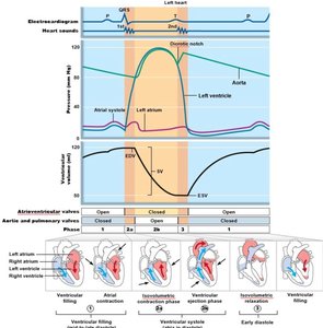
Cardiac Cycle and Wigger’s Diagram
Phases of the Cardiac Cycle
The cardiac cycle describes the sequence of events in one heartbeat, including electrical and mechanical actions. The Wigger’s diagram illustrates changes in pressure, volume, heart sounds, and ECG during the cycle.
Atrial systole: Atria contract, push blood into ventricles; ECG: P wave.
Ventricular systole (isovolumetric contraction): Ventricles contract, AV valves close; ECG: QRS complex; Heart sound: S1 (Lub).
Ventricular ejection: Semilunar valves open, blood leaves heart; volume decreases to ESV.
Ventricular diastole (isometric relaxation): Ventricles relax, semilunar valves close; ECG: T wave; Heart sound: S2 (Dub).
Ventricular filling: AV valves open, blood flows passively into ventricles.
Key Concepts
Systole: Contraction phase (squeeze).
Diastole: Relaxation phase.
Electrical events precede mechanical events: ECG changes occur before muscle contraction.
Pressure and volume changes: Blood volume and pressure fluctuate as chambers fill and empty.
Table: Cardiac Cycle Phases and Events
Phase | ECG Wave | Pressure Change | Valve Status | Heart Sound |
|---|---|---|---|---|
Atrial Systole | P wave | Atrial ↑, Ventricular slight ↑ | AV open, Semilunar closed | None |
Ventricular Systole (Isovolumetric) | QRS complex | Ventricular ↑ | All closed | S1 (Lub) |
Ventricular Ejection | -- | Aortic ↑, Ventricular high | Semilunar open | None |
Ventricular Diastole (Isometric) | T wave | Ventricular ↓ | All closed | S2 (Dub) |
Ventricular Filling | -- | Ventricular low | AV open | None |
Additional info: The Wigger’s diagram integrates ECG, heart sounds, pressure, and volume changes for comprehensive understanding of the cardiac cycle.
