 Back
BackEfferent Division: Autonomic and Somatic Motor Control – Study Notes
Study Guide - Smart Notes
 Tailored notes based on your materials, expanded with key definitions, examples, and context.
Efferent Division of the Peripheral Nervous System
Overview
The efferent division of the peripheral nervous system transmits commands from the central nervous system (CNS) to muscles and glands. It is divided into two main subdivisions: the somatic motor neurons, which control skeletal muscles (mostly voluntary), and the autonomic neurons, which control smooth muscle, cardiac muscle, many glands, and some adipose tissue (mostly involuntary).
The Autonomic Division
Subdivisions of the Autonomic Division
The autonomic division is further divided into two branches:
Sympathetic branch – Responsible for the "fight-or-flight" response, preparing the body for action.
Parasympathetic branch – Responsible for the "rest-and-digest" response, promoting maintenance activities and conserving energy.
These branches are anatomically distinguishable and are best differentiated by their activity in various physiological situations.
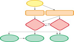
Homeostatic Role of Autonomic Reflexes
Autonomic reflexes are essential for maintaining homeostasis. They work in conjunction with the endocrine and behavioral state systems. Sensory information from somatosensory and visceral receptors, as well as hypothalamic sensors, is integrated in the hypothalamus, pons, and medulla. These centers initiate autonomic, endocrine, and behavioral responses. Some autonomic reflexes are integrated at the spinal cord level (spinal reflexes), bypassing the brain.
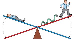
Antagonistic and Cooperative Control
Most internal organs are under antagonistic control, meaning one autonomic branch is excitatory while the other is inhibitory. However, some tissues (e.g., sweat glands, most blood vessels) receive only sympathetic innervation and are under tonic control. In cooperative control, both branches work on different tissues to achieve a common goal. The response in target tissues is often determined by the type of neurotransmitter receptor present.
Autonomic Pathways Structure
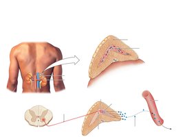
Autonomic pathways consist of two efferent neurons in series:
Preganglionic neuron: Cell body in the CNS, projects to an autonomic ganglion outside the CNS.
Postganglionic neuron: Cell body in the autonomic ganglion, projects to the target tissue.
A ganglion is a cluster of neuronal cell bodies outside the CNS and can act as a mini-integration center.
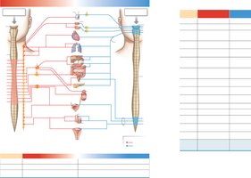
Sympathetic vs. Parasympathetic Anatomy
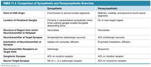
Sympathetic: Originates in thoracic and lumbar regions of the spinal cord; ganglia are located in two chains along the vertebral column.
Parasympathetic: Originates in the brainstem and sacral spinal cord; ganglia are located on or near target organs. The vagus nerve contains about 75% of all parasympathetic fibers.
Chemical Signaling in the Autonomic Nervous System
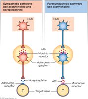
Both sympathetic and parasympathetic preganglionic neurons release acetylcholine (ACh) onto nicotinic cholinergic receptors (nAChR) on postganglionic cells.
Most postganglionic sympathetic neurons secrete norepinephrine (NE) onto adrenergic receptors on target cells.
Most postganglionic parasympathetic neurons secrete ACh onto muscarinic cholinergic receptors (mAChR) on target cells.
Exceptions include sympathetic cholinergic neurons on sweat glands (release ACh) and nonadrenergic, noncholinergic neurons (release other neurotransmitters).
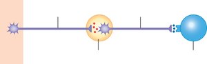
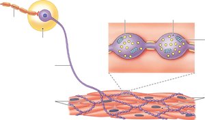
Neuroeffector Junctions and Varicosities
The neuroeffector junction is the synapse between a postganglionic autonomic neuron and its target cell. Varicosities are swellings at the end of postganglionic neurons that release neurotransmitters over the surface of target cells, allowing for widespread effects.
Autonomic Receptor Subtypes
Autonomic receptors are primarily G protein-coupled receptors with multiple subtypes:
Adrenergic receptors (sympathetic):
Alpha (α) receptors: α1 (muscle contraction/secretion), α2 (smooth muscle relaxation/decreased secretion)
Beta (β) receptors: β1 (heart, kidney; equal response to NE and E), β2 (more sensitive to E), β3 (more sensitive to NE)
Muscarinic receptors (parasympathetic): Stimulated by ACh on target cells
Table: Properties of Autonomic Neurotransmitter Receptors
Receptor | Found in | Sensitivity | Effect on Second Messenger |
|---|---|---|---|
α1 | Most sympathetic target tissues | NE > E | Increases IP3 and intracellular Ca2+; increases PKC |
α2 | GI tract and pancreas | NE > E | Decreases cAMP |
β1 | Heart muscle, kidney | NE = E | Increases cAMP |
β2 | Certain blood vessels, smooth muscle | E > NE | Increases cAMP |
β3 | Adipose tissue | NE > E | Increases cAMP |
Muscarinic (M1, M3, M5) | Parasympathetic targets | ACh | Increases IP3 and Ca2+ |
Muscarinic (M2, M4) | Parasympathetic targets | ACh | Decreases cAMP; opens K+ channels |
Table: Comparison of Sympathetic and Parasympathetic Branches
The Adrenal Medulla
The adrenal medulla is a neuroendocrine tissue that acts as a modified sympathetic ganglion. It is innervated by sympathetic preganglionic fibers, and its postganglionic cells (chromaffin cells) lack axons. Instead, they secrete epinephrine (a neurohormone) directly into the blood, allowing for rapid, widespread sympathetic responses.
The Somatic Motor Division
Somatic Motor Pathways
Somatic motor pathways consist of a single neuron originating in the CNS (brain or ventral horn of the spinal cord). These neurons are myelinated, very long, and always excitatory. Each terminal branch innervates a single skeletal muscle fiber. The neuromuscular junction (NMJ) is the synapse between a somatic motor neuron and a skeletal muscle fiber. The motor end plate is the region of the muscle cell membrane that interacts with the neuron, and acetylcholinesterase (AChE) in the synaptic cleft breaks down ACh to terminate the signal.
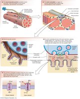
Nicotinic Receptors at the NMJ
The nicotinic acetylcholine receptors (nAChR) at the NMJ are chemically-gated channels with two binding sites for ACh. They are always excitatory, leading to muscle contraction. These receptors are similar to those found on postganglionic autonomic neurons but are specialized for skeletal muscle activation.

Summary Table: Key Differences Between Autonomic and Somatic Motor Divisions
Feature | Autonomic Division | Somatic Motor Division |
|---|---|---|
Number of Neurons | Two (preganglionic and postganglionic) | One |
Target Tissues | Smooth muscle, cardiac muscle, glands, adipose tissue | Skeletal muscle |
Neurotransmitter at Target | NE or ACh | ACh |
Receptor Types | Adrenergic, muscarinic | Nicotinic |
Control | Involuntary | Voluntary |
