 Back
BackEKG Rhythms: Normal and Abnormal Cardiac Conduction
Study Guide - Smart Notes
 Tailored notes based on your materials, expanded with key definitions, examples, and context.
Cardiac Arrhythmias: Overview
Introduction to Cardiac Arrhythmias
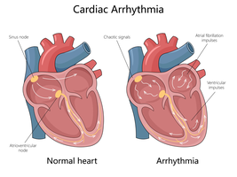
Cardiac arrhythmias are disturbances in the normal rhythm of the heart, often detected and analyzed using an electrocardiogram (EKG or ECG). Understanding these rhythms is essential for recognizing normal cardiac function and diagnosing pathological conditions. This section covers the most common arrhythmias encountered in clinical practice.
Normal Sinus Rhythm
Tachycardia
Bradycardia
Atrial Fibrillation
Inferior Myocardial Infarction (MI)
Atrioventricular (AV) Blocks
Normal Sinus Rhythm
Characteristics of Normal Sinus Rhythm
Normal sinus rhythm is the standard rhythm of a healthy heart, initiated by the sinoatrial (SA) node. It ensures efficient and coordinated contraction of the atria and ventricles.
Heart Rate: 60–100 beats per minute (bpm)
QRS Morphology: Normal (narrow, upright in most leads)
Rhythm: Regular
P Wave: Present before each QRS complex, identical in appearance
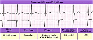
Example: A healthy adult at rest typically exhibits normal sinus rhythm on EKG.
Tachycardia
Sinus Tachycardia
Tachycardia refers to an abnormally fast heart rate, often originating from the SA node. It can be physiological (e.g., exercise, anxiety) or pathological (e.g., fever, anemia).
Heart Rate: Greater than 100 bpm
QRS Morphology: Normal
Rhythm: Regular
P Wave: Present before all beats
Example: Sinus tachycardia may occur during physical exertion or as a compensatory response to hypovolemia.
Bradycardia
Sinus Bradycardia
Bradycardia is defined as a slow heart rate, often due to increased vagal tone or intrinsic disease of the SA node. It is common in athletes and during sleep.
Heart Rate: Less than 60 bpm
QRS Morphology: Normal
Rhythm: Regular
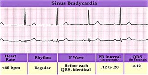
Example: Well-trained athletes may have resting bradycardia without symptoms.
Atrial Fibrillation (A-Fib)
Pathophysiology and EKG Features
Atrial fibrillation is a common arrhythmia characterized by chaotic electrical activity in the atria, leading to ineffective atrial contraction and an irregularly irregular ventricular response.
Rhythm: Irregularly irregular (no predictable pattern)
P Waves: Absent; replaced by fibrillatory ("f") waves that appear as squiggly lines
Ventricular Rate: Can be slow, moderate, or rapid
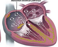
Example: Patients with A-Fib are at increased risk for thromboembolic events such as stroke.
Inferior Myocardial Infarction (MI)
EKG Changes and Clinical Features
An inferior MI is a type of heart attack affecting the lower (inferior) portion of the heart, usually due to occlusion of the right coronary artery. It is diagnosed by characteristic EKG changes.
ST Segment Elevation: Seen in leads II, III, and aVF
Reciprocal ST Depression: May be seen in leads I and aVL
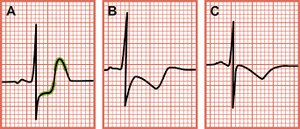
Example: Patients may present with chest pain, hypotension, and bradycardia.
Atrioventricular (AV) Blocks
Overview of AV Blocks
AV blocks are conduction disturbances between the atria and ventricles, classified by the degree of delay or interruption in signal transmission.
First Degree AV Block: Prolonged PR interval, but all atrial impulses are conducted to the ventricles
Second Degree AV Block: Some atrial impulses fail to conduct to the ventricles (subtypes: Mobitz I and II)
Third Degree AV Block: Complete dissociation between atrial and ventricular activity
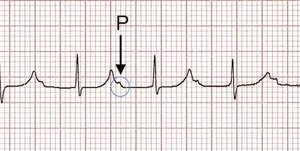
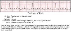
First Degree AV Block
Characterized by a consistently prolonged PR interval (>0.20 seconds) with all P waves followed by QRS complexes.
Rhythm: Regular
PR Interval: Prolonged
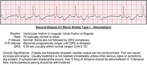
Second Degree AV Block (Mobitz Type I – Wenckebach)
Progressive prolongation of the PR interval until a QRS complex is dropped. Often benign but can cause symptoms if frequent.
Rhythm: Irregular (due to dropped beats)
PR Interval: Progressively lengthens until a beat is missed
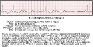
Second Degree AV Block (Mobitz Type II)
Intermittent non-conducted P waves without progressive PR prolongation. More likely to progress to complete block and may require pacing.
Rhythm: Regular or irregular
PR Interval: Constant for conducted beats
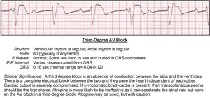
Third Degree (Complete) AV Block
There is no relationship between P waves and QRS complexes; atria and ventricles beat independently. This is a medical emergency requiring immediate intervention.
Rhythm: Regular but dissociated
PR Interval: Variable
Summary Table: Key EKG Features of Common Arrhythmias
Rhythm | Heart Rate | P Wave | QRS | PR Interval |
|---|---|---|---|---|
Normal Sinus | 60–100 bpm | Present, identical | Normal | 0.12–0.20 s |
Tachycardia | >100 bpm | Present | Normal | Normal |
Bradycardia | <60 bpm | Present | Normal | Normal |
Atrial Fibrillation | Variable | Absent (fibrillatory waves) | Normal | Not measurable |
1st Degree AV Block | Usually normal | Present | Normal | >0.20 s |
2nd Degree AV Block I | Usually normal | Present | Normal | Progressively lengthens |
2nd Degree AV Block II | Usually normal | Present | Normal | Constant, some dropped QRS |
3rd Degree AV Block | Usually bradycardic | Present | Normal or wide | Variable, no relationship |
Additional info: Understanding EKG interpretation is crucial for diagnosing and managing cardiac arrhythmias, which can have significant clinical implications ranging from benign to life-threatening conditions.
