 Back
BackElectrical and Mechanical Events of the Heart: Cardiac Physiology and Regulation
Study Guide - Smart Notes
 Tailored notes based on your materials, expanded with key definitions, examples, and context.
Electrical Events of the Heart
Intrinsic Conduction System
The heart's rhythmic contractions are coordinated by the intrinsic conduction system, a network of noncontractile (autorhythmic) cells that initiate and distribute electrical impulses. This system ensures the heart beats in a coordinated manner, independent of direct nervous system stimulation, though it can be modulated by the autonomic nervous system.
Gap junctions allow electrical signals to pass rapidly between cardiac muscle cells.
The intrinsic cardiac conduction system consists of pacemaker cells that generate and propagate action potentials.
Pacemaker Cells and Action Potentials
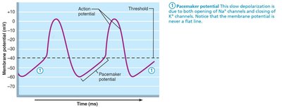
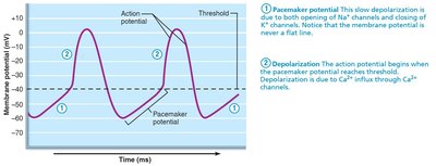
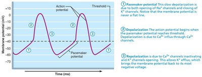
Pacemaker cells, primarily located in the sinoatrial (SA) node, have unstable resting membrane potentials, known as pacemaker potentials or prepotentials. Their action potential consists of three main phases:
Pacemaker potential: Slow depolarization due to opening of Na+ channels and closing of K+ channels, causing the membrane potential to become more positive.
Depolarization: When the threshold is reached, Ca2+ channels open, allowing a rapid influx of Ca2+ and generating the rising phase of the action potential.
Repolarization: Ca2+ channels inactivate and K+ channels open, allowing K+ efflux, which returns the membrane potential to its most negative value.
Sequence of Excitation in the Heart
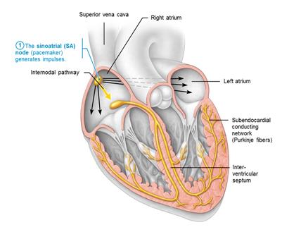
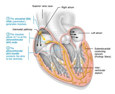
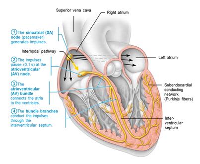
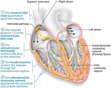
The sequence of excitation ensures the heart contracts in a coordinated manner. The impulse travels through the following structures in approximately 0.22 seconds:
Sinoatrial (SA) node: Pacemaker of the heart, located in the right atrial wall. It generates impulses about 75 times per minute (sinus rhythm).
Atrioventricular (AV) node: Located in the inferior interatrial septum, it delays the impulse by about 0.1 seconds, allowing atrial contraction before ventricular contraction.
Atrioventricular (AV) bundle (Bundle of His): Only electrical connection between atria and ventricles, located in the superior interventricular septum.
Right and left bundle branches: Pathways in the interventricular septum that carry impulses toward the apex of the heart.
Subendocardial conducting network (Purkinje fibers): Completes the pathway through the interventricular septum into the apex and ventricular walls, causing ventricular contraction from apex toward atria.
Autonomic Innervation of the Heart
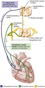
The autonomic nervous system (ANS) modulates heart rate and force of contraction:
Cardioacceleratory center: Sympathetic stimulation increases heart rate and contractility.
Cardioinhibitory center: Parasympathetic stimulation (via the vagus nerve) decreases heart rate.
Action Potentials of Contractile Cardiac Muscle Cells
Phases of Action Potential
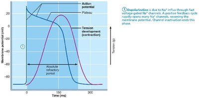
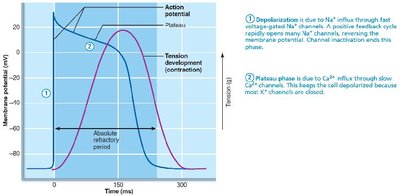
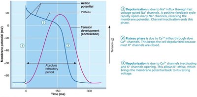
Contractile cardiac muscle fibers are responsible for the heart's pumping action. Their action potential differs from skeletal muscle due to the presence of a plateau phase:
Depolarization: Fast voltage-gated Na+ channels open, causing a rapid influx of Na+ and a sharp rise in membrane potential.
Plateau phase: Slow Ca2+ channels open, maintaining depolarization as most K+ channels remain closed.
Repolarization: Ca2+ channels inactivate and K+ channels open, allowing K+ efflux and returning the membrane potential to resting levels.
The longer action potential and contraction prevent tetanic contractions, ensuring efficient blood ejection.
Electrocardiography (ECG/EKG)
Principles of ECG
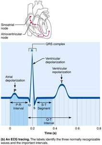
An electrocardiogram (ECG or EKG) is a graphic recording of the heart's electrical activity, representing the sum of all action potentials at a given time. Electrodes are placed at various points on the body to measure voltage differences, typically using a 12-lead system.
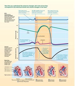
P wave: Depolarization of the SA node and atria.
QRS complex: Ventricular depolarization and atrial repolarization.
T wave: Ventricular repolarization.
P-R interval: Beginning of atrial excitation to beginning of ventricular excitation.
S-T segment: Entire ventricular myocardium depolarized.
Q-T interval: Beginning of ventricular depolarization through ventricular repolarization.
Normal and Abnormal ECG Patterns
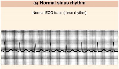
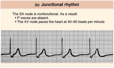
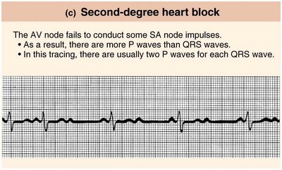
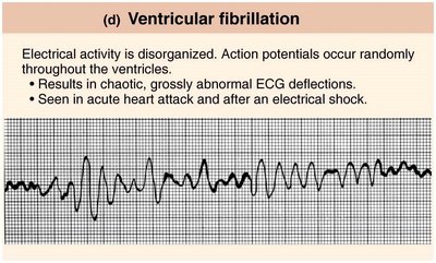
ECG tracings can reveal normal sinus rhythm as well as various arrhythmias and conduction abnormalities:
Normal sinus rhythm: Regular pattern with all waves present.
Junctional rhythm: SA node nonfunctional; P waves absent; AV node paces heart at 40–60 bpm.
Second-degree heart block: AV node fails to conduct some SA node impulses; more P waves than QRS complexes.
Ventricular fibrillation: Disorganized electrical activity; chaotic ECG deflections.
Mechanical Events of the Heart: The Cardiac Cycle
Phases of the Cardiac Cycle
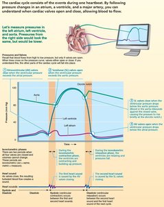
The cardiac cycle describes the sequence of mechanical events during one heartbeat, including periods of contraction (systole) and relaxation (diastole):
Ventricular filling (mid-to-late diastole): Blood flows passively from atria to ventricles; atrial contraction delivers the final 20% of blood (end diastolic volume, EDV).
Isovolumetric contraction: Ventricles contract with all valves closed; pressure rises until semilunar valves open.
Ventricular ejection: Blood is expelled into the aorta and pulmonary trunk.
Isovolumetric relaxation (early diastole): Ventricles relax, semilunar valves close, and the cycle repeats.
Heart Sounds
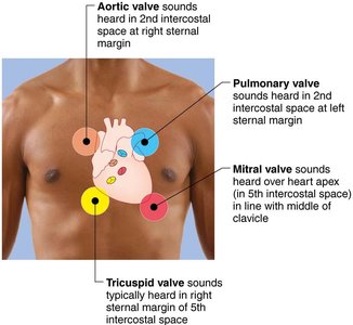
Two main heart sounds ("lub-dup") are associated with valve closures:
First sound (lub): Closing of AV valves at the beginning of ventricular systole.
Second sound (dup): Closing of semilunar valves at the beginning of ventricular diastole.
Heart sounds can be auscultated at specific thoracic locations for each valve.
Regulation of Cardiac Output
Cardiac Output (CO)
Cardiac output is the amount of blood pumped by each ventricle in one minute:
Formula:
At rest: beats/min, ml/beat, L/min
Cardiac reserve is the difference between resting and maximal CO.
Regulation of Stroke Volume (SV)
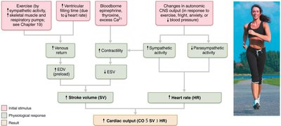
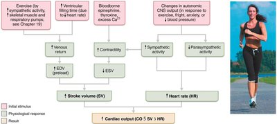
Stroke volume is determined by:
Preload: Degree of stretch of cardiac muscle before contraction (Frank-Starling law).
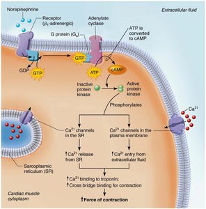
Contractility: Contractile strength at a given muscle length, influenced by sympathetic stimulation and inotropic agents.
Afterload: Pressure that ventricles must overcome to eject blood (mainly arterial pressure).
Mathematically:
Regulation of Heart Rate (HR)
Heart rate is regulated by:
Autonomic nervous system: Sympathetic stimulation increases HR; parasympathetic stimulation decreases HR (vagal tone).
Chemicals: Hormones (epinephrine, thyroxine) and ions (Ca2+, K+) affect HR.
Other factors: Age, gender, exercise, and body temperature.
Clinical Considerations and Homeostatic Imbalances
Arrhythmias: Irregular heart rhythms due to defects in the conduction system.
Heart block: Impaired conduction between atria and ventricles; may require artificial pacemaker.
Congestive heart failure (CHF): Inadequate cardiac output due to weakened myocardium, often from coronary atherosclerosis, hypertension, or myocardial infarcts.
Heart murmurs: Abnormal sounds indicating valve problems (incompetent or stenotic valves).
Developmental Aspects of the Heart
The heart develops from mesoderm, beginning as a simple tube that forms four chambers by day 35 of embryonic development. Fetal adaptations (foramen ovale, ductus arteriosus) bypass pulmonary circulation and close at birth. Age-related changes include valve sclerosis, decreased cardiac reserve, fibrosis, and atherosclerosis.
