 Back
Electrophysiology and Chemical Synaptic Communication in the Nervous System
Study Guide - Smart Notes
 Tailored notes based on your materials, expanded with key definitions, examples, and context.
Nervous System: Electrophysiology and Synaptic Communication
Electrophysiology: Cellular Electrical Phenomena
Electrophysiology is the study of the electrical properties of biological cells and tissues, focusing on the movement of ions across cell membranes and the resulting changes in membrane potential. These electrical events are fundamental to the function of neurons and muscle cells.
Resting Potential (Membrane Potential): The voltage difference across the cell membrane when the cell is at rest, typically around -70 mV in neurons. This is maintained by ion gradients and selective permeability of the membrane.
Action Potential: A rapid, transient change in membrane potential that propagates along the axon, allowing for long-distance communication within the nervous system.
Graded Potential: Local changes in membrane potential that vary in size and decay with distance from the stimulus site.
Phases of Membrane Potential Changes
Depolarization: The membrane potential becomes less negative (moves toward zero), usually due to Na+ influx.
Hyperpolarization: The membrane potential becomes more negative than the resting potential, often due to K+ efflux or Cl- influx.
Repolarization: The return of the membrane potential to resting levels after depolarization, primarily due to K+ efflux.
Types of Electrical Events
Action Potential: All-or-none, long-distance electrical signal.
Graded Potential: Local, variable-strength signal.
Postsynaptic Potential (PSP): Change in membrane potential in the postsynaptic neuron after neurotransmitter binding.
IPSP (Inhibitory Postsynaptic Potential): Hyperpolarizing PSP, decreases likelihood of action potential.
EPSP (Excitatory Postsynaptic Potential): Depolarizing PSP, increases likelihood of action potential.
Ion Channels and Resting Potential
Na+ Leak Channels: Allow slow influx of Na+, slightly depolarizing the cell.
K+ Leak Channels: Allow K+ to exit the cell, making the inside more negative.
Na+/K+ ATPase: Pumps 3 Na+ out and 2 K+ in, maintaining ion gradients and resting potential.
Action Potential Phases and Channel States
Depolarization: Opening of voltage-gated Na+ channels, Na+ influx.
Repolarization: Closing of Na+ channels, opening of voltage-gated K+ channels, K+ efflux.
Hyperpolarization: K+ channels remain open longer, membrane potential becomes more negative than resting.
All-or-None Principle
An action potential either occurs fully or not at all, depending on whether the threshold is reached.
Refractory Periods
Absolute Refractory Period: No new action potential can be generated due to inactivation of Na+ channels.
Relative Refractory Period: A stronger-than-normal stimulus can initiate another action potential; K+ channels are still open, and Na+ channels are resetting.
Action Potential Frequency and Stimulus Intensity
Stronger stimuli increase the frequency of action potentials, not their amplitude.
Speed of Action Potential Conduction
Axon Diameter: Larger diameter = faster conduction.
Myelination: Myelinated axons conduct faster due to saltatory conduction.
Postsynaptic Potentials and Summation
Ligand-Gated Anion Channels: Typically cause hyperpolarization (IPSP).
Ligand-Gated Cation Channels: Typically cause depolarization (EPSP).
Summation: The additive effect of multiple EPSPs and IPSPs at the axon hillock determines if threshold is reached.
Axon Hillock and Neural Integration
The axon hillock integrates all incoming signals (EPSPs and IPSPs) and initiates an action potential if threshold is reached.
Effects of Extracellular Ion Changes
Decreased ECF [K+]: Hyperpolarizes the cell, making action potentials less likely.
Increased ECF [K+]: Depolarizes the cell, making action potentials more likely.
Decreased ECF [Na+]: Reduces action potential amplitude.
Increased ECF [Na+]: Increases action potential amplitude.
Decreased ECF [Cl-]: Reduces hyperpolarization (less inhibitory).
Increased ECF [Cl-]: Increases hyperpolarization (more inhibitory).
Summation of IPSPs and EPSPs: Net effect determines if threshold is reached at the axon hillock.
Demyelinating Diseases
Resting Potential Generation: Usually unaffected.
Action Potential Generation: May be impaired if axonal damage occurs.
Action Potential Propagation: Severely slowed or blocked due to loss of saltatory conduction.
Chemical Synaptic Communication
Key Terms
Neurotransmitter: Chemical messenger released by neurons to transmit signals across a synapse.
Vesicle: Membrane-bound sac storing neurotransmitters in the presynaptic terminal.
Exocytosis: Process by which vesicles release their contents into the synaptic cleft.
Receptor: Protein on the postsynaptic membrane that binds neurotransmitters and initiates a response.
Synaptic Structure
Presynaptic Membrane: The axon terminal membrane that releases neurotransmitters.
Synaptic Cleft: The small gap between presynaptic and postsynaptic membranes.
Postsynaptic Membrane: The membrane of the receiving cell with neurotransmitter receptors.
Voltage-Gated Channels in Presynaptic Membranes
Voltage-gated Na+ channels
Voltage-gated K+ channels
Voltage-gated Ca2+ channels
Postsynaptic Cell Types
Neuron
Muscle cell
Gland cell
Cardiac muscle cell
Smooth muscle cell
Sequence of Chemical Synaptic Communication
Action potential arrives at the presynaptic terminal.
Voltage-gated Ca2+ channels open; Ca2+ enters the terminal.
Ca2+ triggers vesicle fusion and neurotransmitter release (exocytosis).
Neurotransmitter diffuses across the synaptic cleft.
Neurotransmitter binds to receptors on the postsynaptic membrane.
Postsynaptic potential is generated (EPSP or IPSP).
Neurotransmitter Removal Methods
Enzymatic degradation (e.g., acetylcholinesterase breaks down acetylcholine).
Reuptake into presynaptic terminal or glial cells.
Analysis of Ion Channels in Neurons
Channel Type | Location | Possible Ions | Effect on Membrane Potential |
|---|---|---|---|
Leak Channel (A) | Throughout neuron membrane | K+, Na+ | Maintains resting membrane potential |
Voltage-Gated Channel (B) | Axon, axon hillock | Na+, K+, Ca2+ | Initiates and propagates action potentials (depolarization/repolarization) |
Ligand-Gated Channel (C) | Dendrites, cell body | Na+, K+, Cl- | Generates EPSPs or IPSPs (depolarization/hyperpolarization) |
Signal-Gated Channel (D) | Various (depends on cell type) | Ca2+, K+, Cl- | Modulates cell activity in response to intracellular signals |
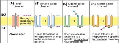
Additional info: The table above summarizes the main types of ion channels, their locations, the ions they conduct, and their effects on membrane potential, as depicted in the provided diagram.
