 Back
Embryonic Development: Key Structures and Processes
Study Guide - Smart Notes
 Tailored notes based on your materials, expanded with key definitions, examples, and context.
Embryonic Development Overview
Introduction
Embryonic development is a fundamental topic in human biology, focusing on the transformation of a fertilized egg into a multicellular organism. This process involves several distinct stages and the formation of specialized structures essential for growth and differentiation.
Stages of Early Embryonic Development
Fertilization and Zygote Formation
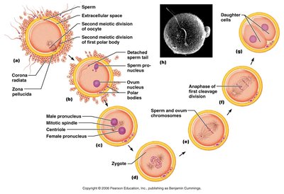
Fertilization occurs when a sperm cell fuses with an ovum, resulting in a zygote. The zygote is the first cell of the new organism and contains a complete set of chromosomes.
Fertilization: Union of male and female gametes.
Zygote: Single cell formed post-fertilization; undergoes rapid mitotic divisions.
Cleavage: Series of mitotic divisions that increase cell number without increasing overall size.
Morula and Blastocyst Formation
After cleavage, the embryo forms a morula (solid ball of cells), which then develops into a blastocyst—a hollow structure with an inner cell mass and an outer trophoblast layer.
Morula: Early solid ball of cells (16-32 cells).
Blastocyst: Fluid-filled cavity forms; consists of trophoblast (outer layer) and inner cell mass (future embryo).
Implantation
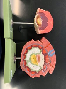
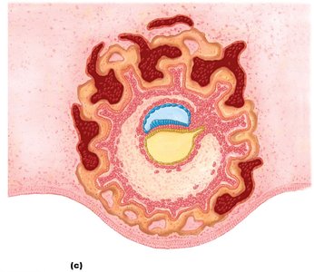
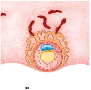
The blastocyst implants into the uterine wall, initiating the formation of extraembryonic structures and the placenta.
Trophoblast: Facilitates implantation and forms part of the placenta.
Inner cell mass: Gives rise to the embryo proper.
Gastrulation and Germ Layer Formation
Gastrulation
Gastrulation is the process by which the blastocyst reorganizes into three primary germ layers: ectoderm, mesoderm, and endoderm. These layers give rise to all tissues and organs.
Epiblast: Source of all three germ layers.
Hypoblast: Contributes to extraembryonic tissues.
Ectoderm: Forms skin, nervous system.
Mesoderm: Forms muscle, bone, blood vessels.
Endoderm: Forms lining of digestive and respiratory tracts.
Major Derivatives of Germ Layers
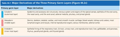
The three primary germ layers differentiate into specific tissues and organs. The following table summarizes their major derivatives:
Primary germ layer | Major derivatives |
|---|---|
Ectoderm (outermost layer) | Epidermis and accessory skin structures, nervous system and organs of the special senses, epithelium of the oral cavity, nasal cavity, and anal canal; adrenal medulla, pituitary, and pineal glands |
Mesoderm (middle layer) | Dermis, skeleton, skeletal, cardiac, and most smooth muscle; cartilage, blood vessels; kidney and ureters; most lymphoid organs and tissues; internal reproductive organs; serous membranes; adrenal cortex |
Endoderm (innermost layer) | Epithelial lining of the respiratory tract, GI tract, urinary tract, and reproductive tract; liver, gallbladder, and pancreas; thymus, parathyroid glands, and thyroid glands |
Extraembryonic Structures
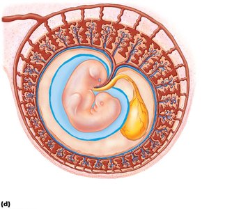
Chorion, Amnion, Yolk Sac, and Allantois
Several extraembryonic membranes form to support the developing embryo:
Chorion: Outermost membrane; forms part of the placenta.
Amnion: Encloses the amniotic cavity, providing a protective fluid environment.
Yolk sac: Early site of blood cell formation.
Allantois: Contributes to the formation of the umbilical cord and urinary bladder.
Placenta and Umbilical Cord
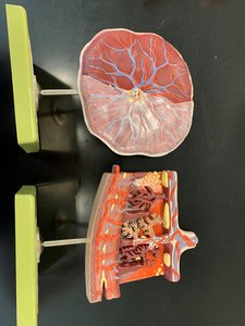
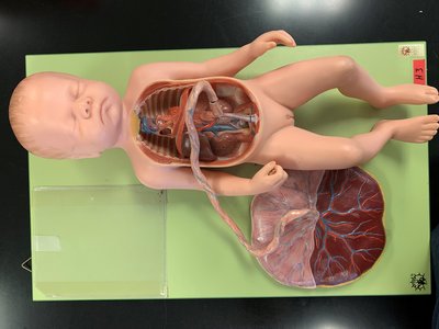
The placenta is a vital organ for nutrient and gas exchange between mother and fetus. The umbilical cord contains two arteries and one vein, connecting the fetus to the placenta.
Chorionic villi: Finger-like projections that increase surface area for exchange.
Decidua basalis: Maternal portion of the placenta.
Intervillous spaces: Areas where maternal blood flows for exchange.
Umbilical cord: Contains two arteries (carry deoxygenated blood from fetus) and one vein (carries oxygenated blood to fetus).
Embryonic and Fetal Development
Progression from Embryo to Fetus
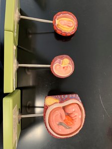
As development continues, the embryo undergoes organogenesis and becomes a fetus. The placenta and membranes continue to support growth and maturation.
Organogenesis: Formation of organs from germ layers.
Fetal period: Growth and functional maturation of organs.
Summary Table: Key Structures and Functions
Structure | Function |
|---|---|
Trophoblast | Implantation, placenta formation |
Inner cell mass | Forms embryo |
Hypoblast | Extraembryonic tissues |
Epiblast | Source of germ layers |
Ectoderm | Skin, nervous system |
Mesoderm | Muscle, bone, blood vessels |
Endoderm | Digestive, respiratory linings |
Chorion | Placenta formation |
Amnion | Protective fluid environment |
Yolk sac | Blood cell formation |
Allantois | Umbilical cord, bladder |
Chorionic villi | Exchange surface |
Decidua basalis | Maternal placenta |
Intervillous spaces | Maternal blood flow |
Placenta | Nutrient/gas exchange |
Umbilical cord | Fetal-maternal connection |
Key Equations and Concepts
Cell Division and Growth
Cell division during cleavage is mitotic, ensuring genetic consistency:
Mitotic division:
Germ layer differentiation:
Conclusion
Understanding embryonic development is essential for comprehending human growth, differentiation, and the origins of tissues and organs. The stages and structures described above form the foundation for advanced study in anatomy and physiology.
