 Back
BackEndocrine and Cardiovascular System Study Guide
Study Guide - Smart Notes
 Tailored notes based on your materials, expanded with key definitions, examples, and context.
Blood: Roles and Components
Functions of Blood
Blood is a vital fluid that performs multiple roles in the human body, including transportation, defense, and homeostasis.
Transportation: Delivers oxygen, nutrients, hormones, and removes metabolic wastes.
Defense: White blood cells (WBCs) and antibodies protect against toxins and pathogens.
Homeostasis: Regulates pH, ion composition, restricts fluid loss at injury sites, and stabilizes body temperature.
Major Components of Blood
Blood consists of plasma and formed elements.
Plasma: 90-92% water, 7% proteins (albumins, globulins, fibrinogen), 1% solutes (nutrients, hormones, electrolytes).
Formed Elements: Erythrocytes (RBCs), leukocytes (WBCs), thrombocytes (platelets).
Red Blood Cells (Erythrocytes)
RBCs are the most numerous formed element and are specialized for oxygen transport.
Contain hemoglobin, which binds oxygen.
Each RBC holds about 300 million hemoglobin molecules.
Hemoglobin types: oxyhemoglobin (with O2), deoxyhemoglobin (without O2), carbaminohemoglobin (with CO2).
White Blood Cells (Leukocytes)
Leukocytes are less numerous than RBCs and are key to immune defense.
Granular: Basophils, Eosinophils, Neutrophils.
Agranular: Monocytes, Lymphocytes.
Mnemonic: "Gran-dpa BEN" for granular types; "Never Let Monkeys Eat Bananas" for abundance order.
Platelets (Thrombocytes)
Platelets are cytoplasmic fragments involved in hemostasis and coagulation.
Derived from megakaryocytes.
Life span: 9–12 days.
Diagnostic Tests and Blood Pathologies
Common Blood Tests
Complete Blood Count (CBC): Measures WBC, RBC, and platelets.
Hemoglobin Concentration: Amount of hemoglobin in blood.
Hematocrit: Percentage of RBCs in total blood volume (W: 37-47%, M: 42-52%).
Blood Disorders
Anemia: Low hematocrit or hemoglobin; includes iron deficiency and sickle cell anemia.
Sickle Cell Anemia: Mutation in hemoglobin gene causes sickle-shaped RBCs.
Hemoglobin Structure and Function
Hemoglobin Components
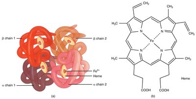
Hemoglobin is a protein complex responsible for oxygen transport in blood.
Composed of four globin chains: alpha 1, alpha 2, beta 1, beta 2.
Each globin is bound to a heme group containing iron.
Example: The heme group binds oxygen via its iron ion, allowing hemoglobin to carry O2 throughout the body.
Leukocyte Types and Disorders
Types of Leukocytes
Neutrophils, lymphocytes, monocytes, eosinophils, basophils.
Granular vs. agranular classification.
Leukocyte Disorders
Leukopenia: Low WBC count.
Leukocytosis: High WBC count.
Leukemia: Cancer affecting leukocytes (lymphocytic or myeloid).
Infectious Mononucleosis: Excessive atypical agranulocytes.
Septicemia: Blood infection.
Lymphoma: Cancer of lymphatic system.
Blood Typing and Compatibility
Blood Group Compatibility
Blood typing determines who can donate or receive blood based on antigens and antibodies.
Universal donor: O-
Universal recipient: AB+
Practice blood group compatibility for safe transfusions.
Blood Clotting Mechanisms
Clotting and Anticoagulants
Platelets and enzymes initiate clotting at injury sites.
Anticoagulants (e.g., protein C, antithrombin, heparin) prevent excessive coagulation.
The Heart: Structure and Function
Heart Chambers
The heart has four chambers, each with a specific role in blood circulation.
Right atrium: Receives blood from vena cava and coronary sinus.
Right ventricle: Pumps blood to pulmonary arteries.
Left atrium: Receives blood from pulmonary veins.
Left ventricle: Pumps blood to aorta.
Heart Valves
Tricuspid (right AV): Right atrium to right ventricle.
Mitral/Bicuspid (left AV): Left atrium to left ventricle.
Pulmonary semilunar: Right ventricle to pulmonary arteries.
Aortic semilunar: Left ventricle to aorta.
Myocardial Layers and Conduction System
Endocardium: Inner layer.
Myocardium: Muscular wall.
Epicardium: Outer layer.
Pericardial cavity: Contains fluid for heart movement.
Fibrous pericardium: Tough outer layer.
Intercalated discs: Connect cardiac muscle cells for synchronized contraction.
Cardiac Cycle
The cardiac cycle consists of systole (contraction) and diastole (relaxation).
Atrial systole: Atria contract, filling ventricles.
Ventricular systole: Ventricles contract, AV valves close, semilunar valves open.
Ventricular diastole: Ventricles relax, semilunar valves close, AV valves open.
Heart Sounds and EKG
Heart Sounds
S1 (lub): AV valves close.
S2 (dub): Semilunar valves close.
Murmurs: Turbulent blood flow, graded 1-6.
Electrocardiogram (EKG)
3, 5, or 12 leads; 12-lead uses 10 electrodes.
P wave: Atrial depolarization.
QRS complex: Ventricular depolarization.
T wave: Atrial repolarization.
Intervals (P-R, QT, ST) indicate heart health.
Cardiac Pathologies
Common Conditions
Angina pectoris: Chest pain.
Myocardial infarction: Heart attack.
Ischemia: Reduced blood supply.
Hypoxia: Low oxygen.
Aneurysm: Arterial bulge.
Atherosclerosis: Plaque buildup.
Cardiac shock: Pump failure.
Cardiac tamponade: Fluid compresses heart.
Endocarditis: Inflammation of inner heart lining.
Pericarditis: Inflammation of pericardium.
Sepsis/Septicemia: Infection and widespread blood infection.
Blood Pressure and Heart Rate
Factors Affecting Blood Pressure
Blood volume, cardiac output, peripheral resistance, compliance.
Hypervolemia: Excess fluid volume.
Baroreceptors: Stretch receptors regulating BP.
Heart Rate Conditions
Bradycardia: HR < 60 BPM.
Tachycardia: HR > 100 BPM.
Normal adult HR: 60-100 BPM; BP: 120/80 mmHg.
Endocrine System Overview
Endocrine vs. Exocrine Glands
Endocrine: Secrete hormones into bloodstream.
Exocrine: Secrete substances directly to target tissue.
Major Endocrine Glands
Pituitary: Master gland; controls other glands.
Thyroid: Regulates metabolism.
Parathyroid: Regulates calcium.
Adrenal: Medulla (epinephrine), cortex (steroids).
Pancreas: Both endocrine (insulin, glucagon) and exocrine.
Testes/Ovaries: Sex hormones.
Pineal: Melatonin, circadian rhythm.
Hematopoiesis
Blood Cell Formation
Occurs in red bone marrow.
Erythropoiesis: RBC production; stimulated by hypoxia.
Leukopoiesis: WBC production; initiated by cytokines.
Thrombopoiesis: Platelet production; initiated by injury.
Platelet Disorders
Common Platelet Disorders
Hemophilia: Missing clotting factor.
Thrombus: Clot in unbroken vessel.
Embolus: Floating clot.
Thrombocytopenia: Low platelets.
Impaired liver function: Vitamin K deficiency.
DIC: Widespread clotting, uncontrolled bleeding.
Cardiac Muscle and Conduction
Structure of Cardiac Muscle
Uni-nucleate, striated, metabolically active.
Intercalated discs: Gap junctions for synchronized contraction.
Conduction System
SA node: Pacemaker.
AV node: Delivers stimulus to Bundle of His.
Bundle branches: Left and right, extend to apex.
Purkinje fibers: Trigger ventricular contraction.
Cardiac Output
CO = HR × SV
Factors: HR (hormones, fitness, age), SV (heart size, contractility, preload, afterload).
Equation:
Additional info: Congenital heart defects and EKG abnormalities (A Fib, V Fib) should be reviewed in textbook for further detail.
