 Back
BackEndocrine and Reproductive Systems: Structure and Function Study Guide
Study Guide - Smart Notes
 Tailored notes based on your materials, expanded with key definitions, examples, and context.
Endocrine System
Overview of the Endocrine System
The endocrine system is a collection of glands that secrete hormones directly into the bloodstream to regulate various body functions. These hormones control processes such as growth, metabolism, and reproduction.
Hypothalamus: Controls the pituitary gland and links the nervous and endocrine systems.
Infundibulum: Connects the hypothalamus to the pituitary gland.
Pituitary Gland: Regulates growth and controls other endocrine glands.
Posterior Pituitary (Pars Nervosa): Secretes ADH and oxytocin.
Anterior Pituitary (Pars Distalis): Secretes GH, TSH, ACTH, LH, FSH, PRL.
Pars Intermedia: Secretes MSH.
Pineal Gland: Secretes melatonin, regulating sleep cycles.
Thyroid Gland: Produces hormones that regulate metabolism, body heat, and bone growth.
Parathyroid Glands: Produce PTH, which raises blood calcium levels.
Thymus Gland: Secretes thymosin for T-cell maturation.
Pancreas: Both endocrine (insulin, glucagon) and exocrine (digestive enzymes) functions.
Kidney: Secretes renin and makes calcitriol from vitamin D.
Adrenal Gland: Cortex secretes aldosterone, cortisol, and androgens; medulla secretes epinephrine and norepinephrine.
Heart: Secretes ANP to lower blood pressure.
Ovary: Secretes estrogen and progesterone.
Testis: Secretes testosterone.
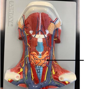
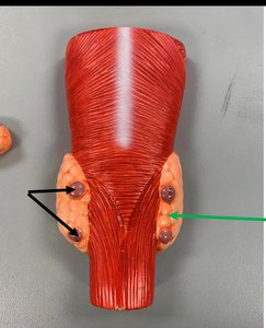
Thyroid and Parathyroid Glands
The thyroid gland is located in the neck and is responsible for producing hormones that regulate metabolism. The parathyroid glands are small glands located on the posterior surface of the thyroid and regulate calcium levels in the blood.
Thyroid Follicle: Made of follicular cells, produces thyroid hormones (T3/T4).
Colloid: Storage of thyroid hormones.
Parafollicular Cells: Produce calcitonin, which lowers blood calcium.
Parathyroid Glands: Produce parathyroid hormone (PTH), which increases blood calcium.
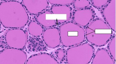
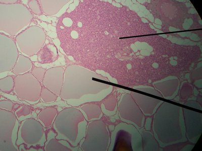
Microscopic Structure of the Thyroid Gland
The thyroid gland is composed of spherical follicles filled with colloid, surrounded by follicular cells. Parafollicular cells are found between follicles.
Follicular Cells: Produce thyroid hormones (T3 and T4).
Colloid: Contains thyroglobulin, a precursor to thyroid hormones.
Parafollicular Cells (C cells): Secrete calcitonin.
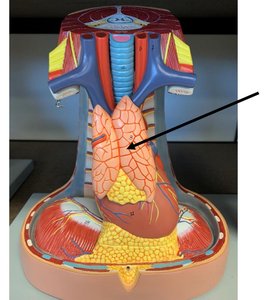
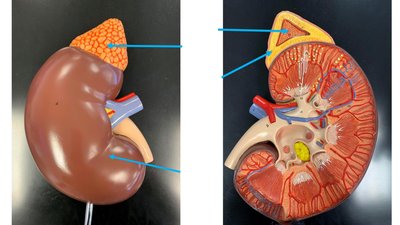
Adrenal Glands
The adrenal glands are located on top of the kidneys and consist of an outer cortex and inner medulla. Each region produces different hormones essential for stress response, metabolism, and electrolyte balance.
Adrenal Cortex: Three zones—zona glomerulosa (aldosterone), zona fasciculata (cortisol), zona reticularis (androgens).
Adrenal Medulla: Secretes epinephrine and norepinephrine.
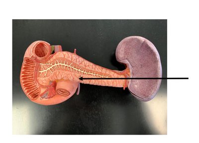
Pancreas
The pancreas has both endocrine and exocrine functions. The endocrine portion consists of pancreatic islets that secrete insulin and glucagon to regulate blood glucose levels.
Pancreatic Islets (Islets of Langerhans): Secrete insulin (lowers blood glucose) and glucagon (raises blood glucose).
Acinar Cells: Secrete digestive enzymes (exocrine function).
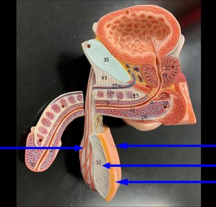
Male Reproductive System
Overview of Male Reproductive Anatomy
The male reproductive system is specialized for the production, storage, and delivery of sperm. It includes both external and internal structures.
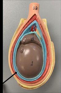
Scrotum: Sac that holds and regulates the temperature of the testes.
Testes: Site of sperm production.
Epididymis: Stores sperm as they mature.
Vas Deferens: Transports sperm from the epididymis to the urethra.
Accessory Glands: Prostate, seminal vesicles, and bulbourethral glands add fluids to semen.
Penis: Organ for delivering sperm during intercourse.
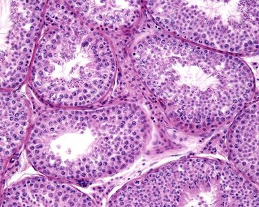
Microscopic Structure of the Testis
The testis contains seminiferous tubules where spermatogenesis occurs. Sertoli cells support developing sperm, and Leydig cells produce testosterone.
Spermatogenesis: Process of sperm production in seminiferous tubules.
Sertoli Cells: Support and nourish developing sperm cells.
Leydig Cells: Located in the interstitial tissue, secrete testosterone.
Female Reproductive System
Overview of Female Reproductive Anatomy
The female reproductive system is specialized for the production of eggs, fertilization, and support of fetal development. It includes both external and internal structures.
Vulva: External genitalia.
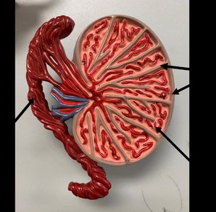
Ovaries: Produce eggs and secrete hormones.
Uterus: Site of implantation and fetal development.
Fallopian Tubes: Conduct eggs from ovaries to uterus; site of fertilization.
Vagina: Muscular passageway for intercourse and childbirth.
Mammary Glands: Produce milk for nourishment of infants.
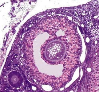
Microscopic Structure of the Ovary
The ovary contains follicles at various stages of development. Each follicle contains an oocyte surrounded by granulosa cells.
Primordial Follicle: Primary oocyte surrounded by a single layer of cells.
Primary Follicle: Immature oocyte with a single layer of granulosa cells.
Secondary Follicle: Multiple layers of granulosa cells and an antrum.
Tertiary (Graafian) Follicle: Large antrum, ready for ovulation.
Corpus Luteum: Secretes progesterone after ovulation.
Reproductive Physiology
Spermatogenesis and Oogenesis
These are the processes by which gametes (sperm and eggs) are produced in males and females, respectively.
Spermatogenesis: Production of sperm in the seminiferous tubules.
Spermiogenesis: Maturation of spermatids into spermatozoa.
Oogenesis: Development of oocytes in the ovary.
Ovulation: Release of a mature ovum from the ovary.
Embryology
Early Developmental Stages
Embryology covers the stages from fertilization to the formation of the basic body plan. Key stages include cleavage, blastula, gastrulation, and organogenesis.
Fertilization: Union of sperm and ovum.
Cleavage: Rapid cell division forming blastomeres.
Morula: Solid ball of cells.
Blastula/Blastocyst: Hollow ball of cells with inner cell mass and trophoblast.
Implantation: Embedding of blastocyst in uterine lining.
Gastrulation: Formation of three germ layers: ectoderm, mesoderm, endoderm.
Placenta: Organ that nourishes and maintains the fetus.
Additional info: The images included are only those that directly reinforce the anatomical and histological structures described in the text, as per the strict relevance requirement.
