 Back
Endocrine System: Structure, Function, and Clinical Aspects
Study Guide - Smart Notes
 Tailored notes based on your materials, expanded with key definitions, examples, and context.
Endocrine System Overview
General Function and Major Organs
The endocrine system is one of the body's two major control systems, interacting with the nervous system to coordinate and integrate the activity of most body cells. It uses hormones—chemical messengers transported in the blood—to influence metabolic activities.
Major processes: Reproduction, growth and development, homeostasis, regulation of cellular metabolism and energy balance, mobilization of body defenses.
Major organs: Pituitary, thyroid, parathyroid, adrenal, pineal glands, hypothalamus (neuroendocrine organ), pancreas, gonads, placenta, and other tissues (heart, kidneys, skin, thymus, bone, adipose).
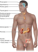
Comparison of Nervous and Endocrine Systems
Nervous system: Fast, uses electrical impulses, effects are short-lived.
Endocrine system: Slow, uses hormones, effects are longer-lasting and widespread.
Hormones and Chemical Messengers
Types of Chemical Messengers
Hormones: Long-distance chemical signals; travel in blood to reach target cells.
Autocrines: Affect the same cells that secrete them.
Paracrines: Affect neighboring cells locally.
Chemical Structure and Classes of Hormones
The chemical structure of a hormone determines its solubility in water, transport, degradation, and receptor interaction.
Amino acid–based hormones: Water soluble (except thyroxine), cannot cross plasma membrane.
Steroid hormones: Synthesized from cholesterol, lipid soluble, can cross plasma membrane.
Eicosanoids: Paracrines and autocrines.
Mechanisms of Hormone Action
Second Messenger Systems
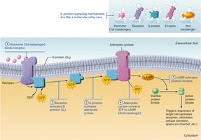
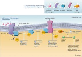
Water-soluble hormones act on plasma membrane receptors and are often coupled via G proteins to second messengers.
cAMP mechanism: Hormone binds receptor → G protein activation → adenylate cyclase activation → ATP converted to cAMP → cAMP activates protein kinases.
PIP2-Calcium mechanism: Hormone activates phospholipase C → splits PIP2 into DAG and IP3 → DAG activates protein kinases, IP3 releases Ca2+ from storage.
Direct Gene Activation
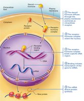
Lipid-soluble hormones (steroids and thyroid hormone) diffuse into cells and bind intracellular receptors, directly activating genes.
Central Dogma: DNA → mRNA → protein.
Proteins synthesized have metabolic, structural, or extracellular functions.
Regulation of Hormone Release
Types of Stimuli
Hormone release is controlled by negative feedback and triggered by three types of stimuli:
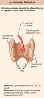
Humoral: Changing blood levels of ions/nutrients (e.g., Ca2+ stimulates PTH release).
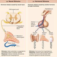
Neural: Nerve fibers stimulate hormone release (e.g., sympathetic fibers stimulate adrenal medulla).
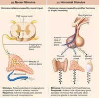
Hormonal: Hormones stimulate other endocrine organs (e.g., hypothalamic hormones regulate pituitary).
Upregulation and Downregulation of Receptors
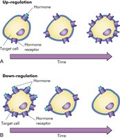
Upregulation: Target cells add receptors in response to low hormone levels, increasing sensitivity.
Downregulation: Cells remove receptors in response to high hormone levels, preventing overstimulation.
Interaction of Hormones at Target Cells
Permissiveness: One hormone requires another to exert its effects.
Synergism: Multiple hormones produce amplified effects.
Antagonism: One hormone opposes the action of another.
Hypothalamus-Pituitary Axis
Structure and Function
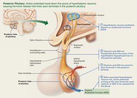
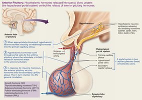
The hypothalamus controls the release of hormones from the pituitary gland via neural and vascular connections.
Posterior pituitary: Neural tissue; stores and secretes oxytocin and ADH.
Anterior pituitary: Glandular tissue; manufactures and secretes six hormones (FLAT PiG: FSH, LH, ACTH, TSH, PRL, GH).
Posterior Pituitary Hormones
Oxytocin: Stimulates uterine contractions and milk ejection; positive feedback mechanism.
Antidiuretic Hormone (ADH): Promotes water reabsorption in kidneys; high concentrations cause vasoconstriction.
Anterior Pituitary Hormones
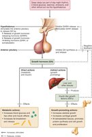
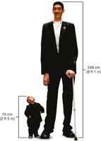
Growth Hormone (GH): Direct actions on metabolism; indirect actions via IGFs stimulate growth of bone and muscle.
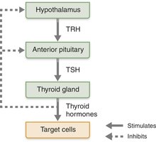
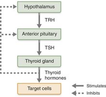
Thyroid-Stimulating Hormone (TSH): Stimulates thyroid gland.
Adrenocorticotropic Hormone (ACTH): Stimulates adrenal cortex.
Follicle-Stimulating Hormone (FSH) & Luteinizing Hormone (LH): Stimulate gonads.
Prolactin (PRL): Stimulates milk production.
Thyroid and Parathyroid Glands
Thyroid Gland
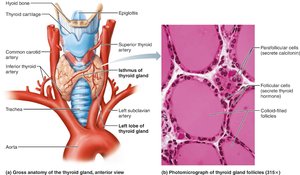
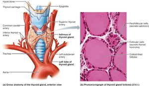
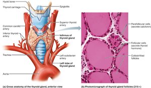
Location: Anterior trachea, inferior to larynx; largest pure endocrine gland.
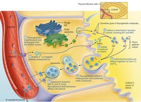
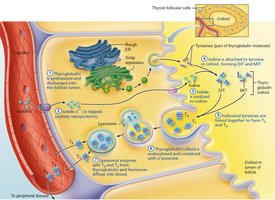
Structure: Follicles produce thyroglobulin; parafollicular cells produce calcitonin.
Hormones: T3 (triiodothyronine) and T4 (thyroxine) regulate metabolism; calcitonin lowers blood calcium.
Clinical Nuggets: Thyroid Disorders
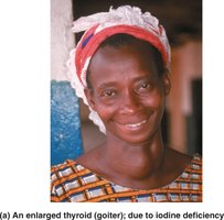
Hypothyroidism: Myxedema, Hashimoto’s thyroiditis, goiter (iodine deficiency).
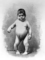
Congenital hypothyroidism (cretinism): Poor development, brain impairment.
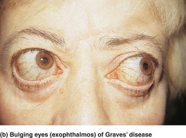
Hyperthyroidism (Graves’ disease): Elevated metabolism, exophthalmos.
Parathyroid Glands
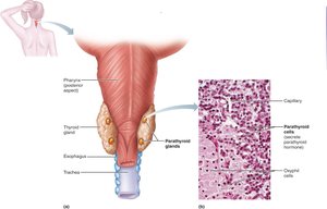
Location: Posterior aspect of thyroid gland; four small glands.
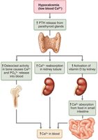
Hormone: Parathyroid hormone (PTH) regulates blood calcium.
Actions: Stimulates osteoclasts, enhances kidney reabsorption of calcium, promotes vitamin D activation for intestinal absorption.
Clinical Nuggets: Parathyroid Disorders
Hyperparathyroidism: Excess PTH, bone demineralization, kidney stones.
Hypoparathyroidism: Insufficient PTH, hypocalcemia, tetany, convulsions.
Adrenal Glands
Structure and Function
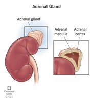
The adrenal glands are pyramid-shaped organs atop the kidneys, consisting of two regions:
Adrenal cortex: Three layers produce corticosteroids.
Adrenal medulla: Produces catecholamines (epinephrine, norepinephrine).
Adrenal Cortex Hormones
Zona glomerulosa: Mineralocorticoids (aldosterone) regulate sodium and potassium.
Zona fasciculata: Glucocorticoids (cortisol) regulate metabolism and stress response.
Zona reticularis: Gonadocorticoids (androgens).
Clinical Nuggets: Adrenal Disorders
Hyperaldosteronism: Excess aldosterone, hypertension, edema.
Cushing’s syndrome: Excess glucocorticoids, high blood glucose, muscle loss.
Addison’s disease: Deficits in corticosteroids, weight loss, dehydration.
Congenital adrenal hyperplasia: Excess androgens, masculinization.
Adrenal Medulla
Catecholamines: Epinephrine and norepinephrine, fight-or-flight response.
Clinical: Pheochromocytoma (tumor), uncontrolled sympathetic activity.
Pineal Gland
Melatonin Secretion
Location: Roof of third ventricle in diencephalon.
Hormone: Melatonin, regulates sleep-wake cycles, antioxidant, antigonadotropic in children.
Pancreas
Structure and Function
The pancreas is a mixed gland with both exocrine and endocrine functions.
Acinar cells: Exocrine, produce digestive enzymes.
Islets of Langerhans: Endocrine, alpha cells secrete glucagon, beta cells secrete insulin.
Glucagon and Insulin
Glucagon: Raises blood glucose by stimulating glycogenolysis and gluconeogenesis in the liver.
Insulin: Lowers blood glucose by promoting cellular uptake and storage.
Clinical Nuggets: Diabetes Mellitus and Hyperinsulinism
Diabetes mellitus: Type 1 (autoimmune, insulin deficiency), Type 2 (insulin resistance).
Symptoms: Polyuria, polydipsia, polyphagia, ketoacidosis.
Hyperinsulinism: Excess insulin, hypoglycemia.
Gonads and Placenta
Hormones and Functions
Ovaries: Estrogens, progesterone, inhibin; regulate reproductive development and cycles.
Testes: Testosterone, inhibin; regulate reproductive development and function.
Placenta: Temporary endocrine organ during pregnancy; secretes estrogens, progesterone, hCG.
Hormone Secretion by Other Organs
Examples
Adipose tissue: Leptin, resistin, adiponectin.
Gastrointestinal tract: Gastrin, ghrelin, secretin, CCK.
Heart: ANP, BNP.
Kidneys: Erythropoietin, renin.
Skeleton: Osteocalcin.
Skin: Cholecalciferol (vitamin D precursor).
Thymus: Thymosins, thymulin, thymopoietins.
Developmental Aspects of the Endocrine System
Endocrine Function Throughout Life
GH, estrogen, testosterone, TH, and PTH levels change with age, affecting metabolism, muscle mass, and bone health.
Glucose tolerance declines, increasing risk for type 2 diabetes.
Summary Table: Major Endocrine Glands and Their Hormones
Gland | Hormone(s) | Main Function |
|---|---|---|
Pituitary (anterior) | GH, TSH, ACTH, FSH, LH, PRL | Growth, metabolism, reproduction |
Pituitary (posterior) | Oxytocin, ADH | Uterine contraction, water balance |
Thyroid | T3, T4, Calcitonin | Metabolism, calcium regulation |
Parathyroid | PTH | Calcium homeostasis |
Adrenal cortex | Aldosterone, cortisol, androgens | Electrolyte balance, stress response, sex hormones |
Adrenal medulla | Epinephrine, norepinephrine | Fight-or-flight response |
Pineal | Melatonin | Sleep-wake cycle |
Pancreas | Insulin, glucagon | Blood glucose regulation |
Ovaries | Estrogen, progesterone | Reproduction |
Testes | Testosterone | Reproduction |
