 Back
Endocrine System: Structure, Function, and Regulation
Study Guide - Smart Notes
 Tailored notes based on your materials, expanded with key definitions, examples, and context.
Endocrine System Overview
Structure and Function of the Endocrine System
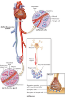
The endocrine system is composed of ductless glands that synthesize and secrete hormones directly into the bloodstream. These hormones travel throughout the body and bind to specific receptors on target cells, eliciting physiological responses. The endocrine system regulates many vital processes, including growth, metabolism, blood composition, digestion, and reproduction.
Hormones: Chemical messengers released by endocrine glands.
Target Cells: Cells with specific receptors for a hormone, enabling them to respond.
Hormone Transport: Hormones are released into interstitial fluid, enter the blood, and bind to target cell receptors.
Comparison of Endocrine and Nervous Systems
The endocrine and nervous systems are the body's two main control systems. Both use ligands (chemical messengers) to communicate, but differ in their methods and effects:
Endocrine System: Transmits hormones via blood, targets any cell with the correct receptor, has widespread effects, slower reaction times, and longer-lasting effects.
Nervous System: Uses neurotransmitters, targets specific cells, has rapid and short-lived effects.
Major Endocrine Glands and Organs
Location of Major Endocrine Glands
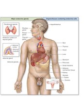
Endocrine glands are distributed throughout the body. Some organs have solely endocrine functions, while others contain clusters of endocrine cells within organs that have other primary functions.
Primary Endocrine Organs: Pituitary, pineal, thyroid, parathyroid, adrenal glands.
Organs with Endocrine Cells: Hypothalamus, skin, thymus, heart, liver, stomach, pancreas, small intestine, adipose tissue, kidneys, gonads.
Hormone Release and Regulation
Stimulation of Hormone Synthesis and Release
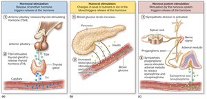
Hormone release is regulated by endocrine reflexes, which can be initiated by hormonal, humoral, or nervous stimuli:
Hormonal Stimulation: Release of one hormone triggers the release of another (e.g., TSH stimulates TH release).
Humoral Stimulation: Changes in blood levels of nutrients or ions trigger hormone release (e.g., increased blood glucose stimulates insulin release).
Nervous System Stimulation: Neuronal signals trigger hormone release (e.g., sympathetic stimulation causes adrenal medulla to release epinephrine).
Hormone Classification
Types of Circulating Hormones
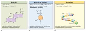
Hormones are classified based on their chemical structure and solubility:
Steroids: Lipid-soluble molecules synthesized from cholesterol (e.g., estrogen, cortisol).
Biogenic Amines: Modified amino acids, mostly water-soluble except thyroid hormone (e.g., norepinephrine, melatonin).
Proteins: Water-soluble chains of amino acids (e.g., insulin, growth hormone).
Hormone Transport and Action
Lipid-Soluble Hormones
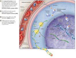
Lipid-soluble hormones (such as steroids and thyroid hormone) require carrier proteins for transport in blood. They diffuse across cell membranes and bind to intracellular receptors, forming a hormone-receptor complex that interacts with DNA to regulate gene expression.
Carrier Proteins: Protect hormones from early destruction; only unbound hormone can exit blood and bind to target cells.
Intracellular Receptors: Located in cytosol or nucleus; hormone-receptor complex binds to hormone-response element (HRE) on DNA, leading to mRNA transcription and protein synthesis.
Water-Soluble Hormones
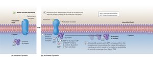
Water-soluble hormones (such as proteins and most biogenic amines) bind to cell-surface receptors and initiate a signal transduction pathway involving G-proteins and second messengers.
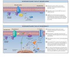
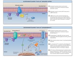
Signal Transduction: Hormone (first messenger) binds to receptor, activates G-protein, which then activates membrane enzymes (adenylate cyclase or phospholipase C).
Second Messengers: cAMP (via adenylate cyclase) or DAG/IP3 (via phospholipase C) modify cellular activity.
Regulation of Hormone Sensitivity and Interactions
Receptor Regulation
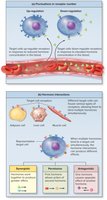
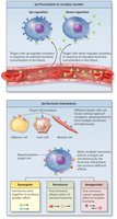
The number of hormone receptors on a target cell can fluctuate, affecting sensitivity:
Up-Regulation: Increase in receptor number, enhancing sensitivity (often occurs when hormone levels are low).
Down-Regulation: Decrease in receptor number, reducing sensitivity (often occurs when hormone levels are high).
Hormone Interactions
Multiple hormones can interact at target cells, producing synergistic, permissive, or antagonistic effects:
Synergistic: Hormones reinforce each other's activity (e.g., estrogen and progesterone).
Permissive: One hormone requires another's activity (e.g., oxytocin requires prolactin).
Antagonistic: Hormones oppose each other's effects (e.g., glucagon vs. insulin).
Hypothalamus and Pituitary Gland
Anatomic Relationship
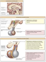
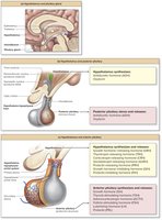
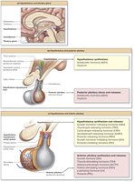
The hypothalamus controls the pituitary gland, which in turn regulates several other endocrine organs. The pituitary is divided into anterior and posterior lobes, each with distinct functions and connections to the hypothalamus.
Posterior Pituitary: Neural connection; stores and releases ADH and oxytocin produced by hypothalamic neurons.
Anterior Pituitary: Vascular connection via the hypothalamo-hypophyseal portal system; releases hormones in response to hypothalamic regulatory hormones.
Anterior Pituitary Hormones
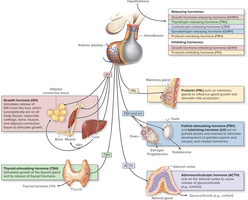
The anterior pituitary releases several key hormones, each regulated by hypothalamic releasing or inhibiting hormones:
Growth Hormone (GH): Stimulates growth and metabolism via IGFs.
Thyroid-Stimulating Hormone (TSH): Stimulates thyroid hormone release.
Adrenocorticotropic Hormone (ACTH): Stimulates adrenal cortex to release corticosteroids.
Prolactin (PRL): Stimulates milk production.
Follicle-Stimulating Hormone (FSH) & Luteinizing Hormone (LH): Regulate gonadal function.
Growth Hormone Regulation and Effects
Regulation of Growth Hormone
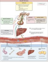
Growth hormone release is regulated by GHRH from the hypothalamus, which stimulates the anterior pituitary. GH stimulates the liver to release IGFs, which together promote growth and nutrient release. Negative feedback regulates this pathway.
Stimuli: Age, time of day, nutrient levels, stress, and exercise.
Effects: Increased protein synthesis, cell division, nutrient release (glycogenolysis, gluconeogenesis, lipolysis).
Thyroid Gland and Hormone Regulation
Anatomy and Function
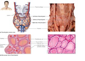
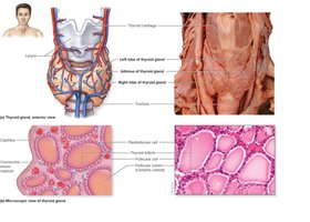
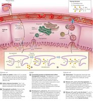
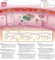
The thyroid gland is located anterior to the trachea and produces thyroid hormone (TH) and calcitonin. Follicular cells synthesize TH, while parafollicular cells produce calcitonin.
TH: Increases metabolic rate, protein synthesis, and energy production.
Calcitonin: Decreases blood calcium levels.
Regulation of Thyroid Hormone
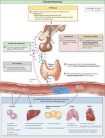
TH release is regulated by TRH from the hypothalamus and TSH from the anterior pituitary. TH increases metabolic rate and is regulated by negative feedback.
Stimuli: Blood levels of TH, environmental factors.
Effects: Increased metabolic rate, glucose-sparing effect, increased heart rate and respiration.
Adrenal Glands and Cortisol Regulation
Anatomy and Function
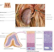
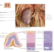
The adrenal glands are located on the superior surface of each kidney and consist of the adrenal cortex and medulla. The cortex produces corticosteroids, including mineralocorticoids, glucocorticoids, and gonadocorticoids.
Mineralocorticoids: Regulate electrolyte levels (e.g., aldosterone).
Glucocorticoids: Regulate blood sugar (e.g., cortisol).
Gonadocorticoids: Sex hormones (e.g., androgens).
Regulation of Cortisol
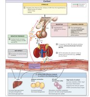
Cortisol release is regulated by CRH from the hypothalamus and ACTH from the anterior pituitary. Cortisol increases blood nutrient levels and is regulated by negative feedback.
Stimuli: Blood levels of cortisol, time of day, stress.
Effects: Increased glycogenolysis, gluconeogenesis, lipolysis, protein catabolism, decreased glucose uptake.
Pancreas and Regulation of Blood Glucose
Anatomy and Function
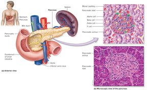
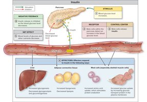
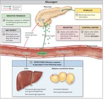
The pancreas has both endocrine and exocrine functions. Pancreatic islets contain alpha cells (secrete glucagon) and beta cells (secrete insulin).
Insulin: Lowers blood glucose by promoting uptake and storage.
Glucagon: Raises blood glucose by promoting release from storage.
Regulation and Action of Insulin
Insulin is released in response to increased blood glucose and promotes nutrient uptake and storage. Negative feedback inhibits further release as glucose levels decrease.
Effects: Increased glycogenesis, lipogenesis, protein synthesis, glucose uptake.
Regulation and Action of Glucagon
Glucagon is released in response to decreased blood glucose and promotes nutrient release into the blood. Negative feedback inhibits further release as glucose levels rise.
Effects: Increased glycogenolysis, gluconeogenesis, lipolysis; decreased glycogenesis, lipogenesis.
Additional Endocrine Structures and Aging
Pineal and Parathyroid Glands
Pineal Gland: Secretes melatonin, regulates circadian rhythm.
Parathyroid Glands: Secrete parathyroid hormone (PTH), increases blood calcium.
Other Endocrine Functions
Thymus: Secretes thymic hormones for T-cell maturation.
Heart: Secretes atrial natriuretic peptide (ANP) to lower blood pressure.
Kidneys: Release erythropoietin (EPO) to increase red blood cell production.
Liver: Secretes IGFs and angiotensinogen.
Stomach/Intestine: Secrete gastrin, secretin, and CCK for digestion.
Adipose Tissue: Secretes leptin to regulate appetite.
Aging and the Endocrine System
Secretory activity of endocrine glands decreases with age, reducing hormone levels and efficiency of endocrine functions. This can lead to loss of body mass and other age-related changes.
Hormone | Source | Main Function |
|---|---|---|
Insulin | Pancreas (Beta cells) | Lowers blood glucose |
Glucagon | Pancreas (Alpha cells) | Raises blood glucose |
Cortisol | Adrenal cortex | Increases blood nutrient levels |
Thyroid Hormone (TH) | Thyroid gland | Increases metabolic rate |
Growth Hormone (GH) | Anterior pituitary | Stimulates growth and metabolism |
Additional info: This guide covers the main concepts and regulatory mechanisms of the endocrine system, suitable for exam preparation in an anatomy and physiology college course.
