Back
Epithelial Tissue: Structure, Types, and Glandular Classification
Study Guide - Smart Notes
 Tailored notes based on your materials, expanded with key definitions, examples, and context.
Epithelial Tissue
Overview of Epithelial Tissue
Epithelial tissue forms the covering or lining of all body surfaces, cavities, and hollow organs. It also constitutes the major tissue in glands. Epithelial cells are closely packed, with minimal extracellular material, and serve as a barrier and interface for absorption, secretion, and protection.
Types of Epithelial Tissue
Simple Columnar Epithelium
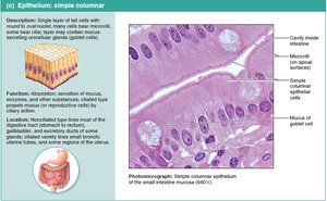
Simple columnar epithelium consists of a single layer of tall, closely packed cells aligned in a columnar fashion. These cells often contain microvilli or cilia and may include mucus-secreting goblet cells.
Description: Single layer of tall cells with round to oval nuclei; some cells bear cilia; layer may contain mucus-secreting unicellular glands (goblet cells).
Functions: Absorption; secretion of mucus, enzymes, and other substances; ciliated type propels mucus (or reproductive cells) by ciliary action.
Location: Nonciliated type lines most of the digestive tract (stomach to rectum), gallbladder, and excretory ducts of some glands; ciliated variety lines small bronchi, uterine tubes, and some regions of the uterus.
Example: Lining of the small intestine.
Pseudostratified Columnar Epithelium
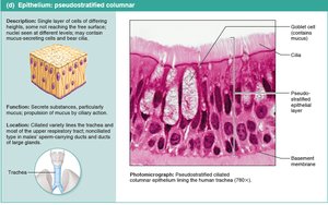
Pseudostratified columnar epithelium appears to have multiple layers due to the varying positions of nuclei, but all cells rest on the basement membrane. It often contains cilia and goblet cells.
Description: Single layer of cells of differing heights; nuclei seen at different levels; may contain mucus-secreting cells and bear cilia.
Functions: Secretion, particularly of mucus; propulsion of mucus by ciliary action.
Location: Ciliated variety lines the trachea and most of the upper respiratory tract; nonciliated type in male sperm-carrying ducts and ducts of large glands.
Example: Lining of the trachea.
Transitional Epithelium
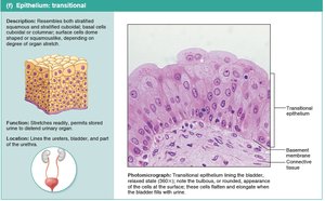
Transitional epithelium is a specialized tissue that can stretch and change shape, appearing cuboidal or squamous depending on the degree of organ distension.
Description: Resembles both stratified squamous and stratified cuboidal; basal cells are cuboidal or columnar; surface cells dome-shaped or squamous-like, depending on degree of organ stretch.
Functions: Stretches readily and permits stored urine to distend urinary organ.
Location: Lines the ureters, bladder, and part of the urethra.
Example: Lining of the urinary bladder.
Glandular Epithelium
Formation and Types of Glands
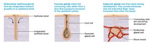
Glandular epithelium forms glands that are specialized for secretion. Glands are classified as endocrine (secreting into the blood) or exocrine (secreting onto body surfaces or into body cavities).
Multicellular exocrine glands develop from invaginations of epithelial sheets and retain connecting ducts to the surface.
Endocrine glands lose their ducts during development and release hormones directly into the bloodstream.
Classification of Exocrine Glands by Structure
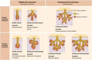
Exocrine glands are classified based on the structure of their ducts (simple or compound) and the shape of their secretory units (tubular or alveolar).
Simple Duct Structure (Duct does not branch) | Compound Duct Structure (Duct branches) | |
|---|---|---|
Tubular secretory structure | Simple tubular Example: Intestinal glands Simple branched tubular Example: Stomach (gastric) glands | Compound tubular Example: Duodenal glands of small intestine |
Alveolar secretory structure | Simple alveolar Example: No important example in humans Simple branched alveolar Example: Sebaceous (oil) glands | Compound alveolar Example: Mammary glands Compound tubuloalveolar Example: Salivary glands |
Modes of Secretion in Exocrine Glands
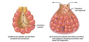
Exocrine glands secrete their products by different mechanisms:
Merocrine secretion: Products are secreted by exocytosis without loss of cellular material. Example: Sweat glands, salivary glands.
Holocrine secretion: The entire secretory cell ruptures, releasing secretions and dead cell fragments. Example: Sebaceous (oil) glands.