 Back
Essential Study Notes: Nervous, Muscular, Cardiovascular, and Sensory Systems
Study Guide - Smart Notes
 Tailored notes based on your materials, expanded with key definitions, examples, and context.
Nervous System
Overview of the Nervous System
The nervous system is a complex network responsible for transmitting signals throughout the body, enabling communication, coordination, and control of bodily functions. It consists of the central nervous system (CNS) and the peripheral nervous system (PNS).
Central Nervous System (CNS): Includes the brain and spinal cord, which process and integrate information.
Peripheral Nervous System (PNS): Composed of nerves that branch out from the CNS to the rest of the body.
Function: Sensory input, integration, motor output.

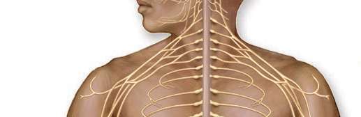
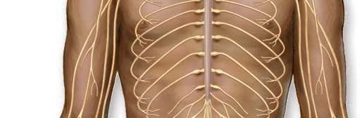
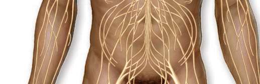
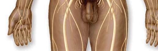
Structure of the Nervous System
The nervous system is organized into distinct regions and pathways, each with specialized functions.
Brain: The control center for processing sensory information and directing responses.
Spinal Cord: Transmits signals between the brain and the rest of the body.
Nerves: Bundles of axons that carry electrical impulses.



Microscopic Structure of Nerves
Nerves are composed of bundles of axons, surrounded by protective layers.
Axon: The long, slender projection of a neuron that conducts electrical impulses.
Myelin Sheath: Insulating layer that increases the speed of impulse transmission.
Endoneurium, Perineurium, Epineurium: Connective tissue layers that protect and organize nerve fibers.



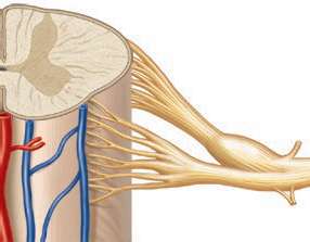
Spinal Cord and Nerve Roots
The spinal cord is a major pathway for information traveling between the brain and peripheral nerves.
Gray Matter: Contains neuron cell bodies.
White Matter: Contains myelinated axons.
Nerve Roots: Dorsal (sensory) and ventral (motor) roots connect the spinal cord to peripheral nerves.

Brain Structure and Pathology
The brain is divided into regions with specialized functions. Pathological changes can be visualized using imaging techniques.
Cerebrum: Responsible for higher functions such as thought and action.
Cerebellum: Coordinates movement and balance.
Imaging: MRI scans can reveal abnormalities such as strokes or tumors.






Muscular System
Structure and Function of Muscles
Muscles are responsible for movement, posture, and heat production. They are composed of muscle fibers that contract in response to nervous stimulation.
Skeletal Muscle: Voluntary muscles attached to bones.
Muscle Fiber: The basic unit of muscle tissue.
Tendon: Connects muscle to bone.

Cardiovascular System
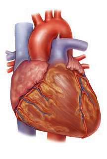
Structure of the Heart
The heart is a muscular organ that pumps blood throughout the body, supplying oxygen and nutrients and removing waste products.
Atria and Ventricles: Chambers that receive and pump blood.
Valves: Ensure unidirectional flow of blood.
Coronary Vessels: Supply blood to the heart muscle itself.
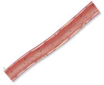
Sensory Systems
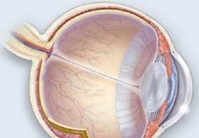
Structure of the Eye
The eye is a specialized organ for vision, converting light into neural signals.
Cornea: Transparent front part of the eye.
Lens: Focuses light onto the retina.
Retina: Contains photoreceptor cells that detect light.

Additional Nervous System Details
Neurons and Glial Cells
Neurons are the functional units of the nervous system, transmitting electrical impulses. Glial cells support and protect neurons.
Neuron: Cell specialized for communication.
Glial Cells: Provide support, nutrition, and insulation.


Peripheral Nerve Organization
Peripheral nerves are organized into fascicles, each surrounded by connective tissue.
Fascicle: Bundle of nerve fibers.
Connective Tissue Layers: Endoneurium, perineurium, epineurium.
Summary Table: Nervous System Components
Component | Structure | Function |
|---|---|---|
Brain | Cerebrum, cerebellum, brainstem | Processing, integration, control |
Spinal Cord | Gray and white matter | Signal transmission |
Peripheral Nerves | Axons, myelin, connective tissue | Signal conduction |
Muscle | Muscle fibers, tendons | Movement |
Heart | Atria, ventricles, valves | Blood circulation |
Eye | Cornea, lens, retina | Vision |
Key Definitions
Neuron: A nerve cell specialized for transmitting electrical impulses.
Axon: The long projection of a neuron that carries signals away from the cell body.
Myelin: Fatty substance that insulates axons and speeds up signal transmission.
Muscle Fiber: The basic unit of muscle tissue, capable of contraction.
Cornea: The transparent front part of the eye.
Ventricle: Lower chamber of the heart that pumps blood out.
Important Equations
Nerve Impulse Transmission
The speed of nerve impulse transmission is influenced by myelination and axon diameter.
Conduction Velocity: where is velocity, is diameter, is membrane resistance, is membrane capacitance.
Cardiac Output
Cardiac output is the volume of blood pumped by the heart per minute.
Formula: where is cardiac output, is heart rate, is stroke volume.
Examples and Applications
Example: Damage to the spinal cord can disrupt communication between the brain and peripheral nerves, leading to loss of sensation or movement.
Application: MRI scans are used to diagnose brain pathologies such as stroke or tumors.
Example: Myelin sheath disorders, such as multiple sclerosis, slow down nerve impulse transmission.
Additional info:
Images and diagrams are inferred to represent anatomical structures relevant to the nervous, muscular, cardiovascular, and sensory systems.
Some images are partial or cropped but are included only when they clearly illustrate the relevant anatomy.
