 Back
BackFinals Review: Human Body Organization, Homeostasis, Cell Structure, and Tissue Fundamentals
Study Guide - Smart Notes
 Tailored notes based on your materials, expanded with key definitions, examples, and context.
The Human Body: An Orientation
Levels of Structural Organization
The human body is organized into a hierarchy of structural levels, each building upon the previous to form the complete organism.
Chemical level: Atoms and molecules form the foundation of all matter.
Cellular level: Cells and their organelles are the basic units of life.
Tissue level: Groups of similar cells work together to perform specific functions.
Organ level: Structures composed of two or more tissue types that perform specific functions.
Organ system level: Organs that work closely together to accomplish a common purpose.
Organismal level: The sum of all organ systems working together to sustain life.

Maintaining Homeostasis
Negative Feedback Loops
Homeostasis is the maintenance of a stable internal environment. Negative feedback loops are the primary mechanisms for maintaining homeostasis, counteracting changes to restore balance.
Receptor: Detects changes in the environment (stimulus).
Control center: Processes information and determines the response.
Effector: Carries out the response to restore homeostasis.
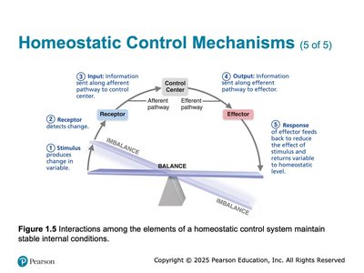
Example: Regulation of body temperature—when body temperature rises, sweat glands (effectors) are activated to cool the body; when temperature drops, muscles shiver to generate heat.

Positive Feedback Loops
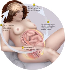
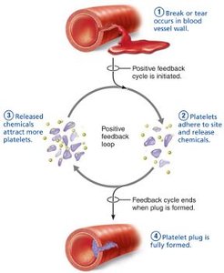
Positive feedback mechanisms amplify the original stimulus, leading to an even greater response. These are less common and typically control infrequent events that require a rapid or definitive outcome.
Response enhances the original stimulus (e.g., labor contractions, blood clotting).
Exhibits a cascade or amplifying effect.
Example 1: Enhancement of labor contractions by oxytocin.
Example 2: Platelet plug formation and blood clotting.
Anatomical Terms and Position
Anatomical Terminology
Standardized anatomical terms are used to describe locations, directions, and movements of the body, ensuring clear communication among healthcare and scientific professionals.
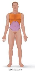
Anatomical position: Body erect, feet slightly apart, palms facing forward, thumbs pointing away from the body.
Importance: Serves as the reference point for describing body parts and regions.
Chemistry Comes Alive
Organic Compounds: Synthesis and Hydrolysis
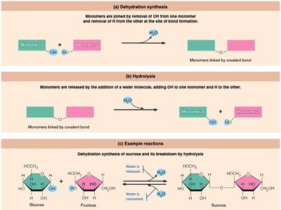
Organic molecules are carbon-based compounds essential for life. They are synthesized and broken down through dehydration synthesis and hydrolysis reactions.
Dehydration synthesis: Monomers are joined by removal of a water molecule, forming covalent bonds.
Hydrolysis: Monomers are released by the addition of a water molecule, breaking covalent bonds.
Major organic compounds: Carbohydrates, lipids, proteins, nucleic acids.

Nucleic Acids: ATP
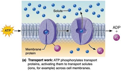
Adenosine triphosphate (ATP) is the primary energy carrier in cells, driving various types of cellular work.
Transport work: ATP phosphorylates transport proteins, enabling movement of substances across membranes.
Mechanical work: ATP provides energy for muscle contraction and movement of cellular structures.
Chemical work: ATP supplies energy for chemical reactions, such as synthesis of macromolecules.

Cells: The Living Units
Cell Surface and Communication
The cell surface is critical for communication, attachment, and transport. Specialized structures and junctions facilitate these functions.
Binding of signaling molecules: Hormones and other signals bind to receptors, triggering cellular responses.
Attachment: Cells adhere to each other and the extracellular matrix.
Transport: Movement of materials into and out of the cell is tightly regulated.
Cell Surface Extensions
Microvilli: Increase surface area for absorption (e.g., small intestine).
Cilia: Move substances across the cell surface (e.g., respiratory tract).
Flagella: Propel cells (e.g., sperm cells).
Pseudopods: Enable cell movement and engulfment of particles.
Cell Junctions
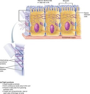
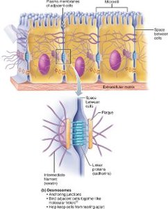
Cell junctions are specialized connections between cells that maintain tissue integrity and facilitate communication.
Tight junctions: Seal adjacent cells to prevent leakage of extracellular fluid (e.g., in the stomach and intestines).

Desmosomes: Anchor cells together, providing mechanical stability (e.g., in the epidermis).
Gap junctions: Allow passage of ions and small molecules for cell-to-cell communication (e.g., in cardiac muscle).
Membrane Transport
Substances move across the plasma membrane by passive or active transport mechanisms.
Passive transport: Does not require energy (e.g., diffusion, osmosis).
Active transport: Requires energy (ATP) to move substances against their concentration gradient.
The Cell Life Cycle
Phases of the Cell Cycle
Cells reproduce by duplicating their DNA and dividing into two daughter cells. The cell cycle consists of interphase and the mitotic phase.
Interphase: Period of growth and DNA replication (includes G1, S, and G2 phases).
Mitotic phase: Division of the nucleus (mitosis) and cytoplasm (cytokinesis).

DNA Replication (S Phase)
During the S phase of interphase, the cell replicates its DNA in preparation for division.
DNA "unzips" and each strand serves as a template for a new complementary strand, synthesized by DNA polymerase.
Result: Two identical sets of DNA for daughter cells.
Mitosis and Cytokinesis
Mitosis is the process by which the nucleus divides, followed by cytokinesis, which divides the cytoplasm, resulting in two genetically identical daughter cells.
Gene Expression: Transcription and Translation
Transcription
Transcription is the process of synthesizing messenger RNA (mRNA) from a DNA template in the nucleus.
mRNA: Carries the genetic code from DNA to the cytoplasm for protein synthesis.
Translation
Translation occurs in the cytoplasm, where ribosomes read the mRNA sequence and assemble amino acids into proteins using transfer RNA (tRNA).
Codon: A sequence of three nucleotides on mRNA that codes for a specific amino acid.
tRNA: Brings the appropriate amino acid to the ribosome based on codon-anticodon pairing.
Tissues: The Living Fabric
Overview of Histology
Histology is the study of tissues and their organization into organs. Tissues are groups of similar cells and their products that perform specific functions.
Four primary tissue types: Epithelial, connective, muscle, and nervous tissue.
Differences: Cell types, functions, matrix characteristics, and the proportion of cells to matrix.
Epithelial Tissue Functions
Protection: Shields underlying tissues from injury and infection (e.g., epidermis).
Secretion: Produces substances such as mucus, sweat, and hormones.
Excretion: Removes waste products (e.g., CO2 across pulmonary epithelium).
Absorption: Takes in nutrients (e.g., small intestine epithelium).
Filtration: Selectively filters substances (e.g., blood vessel endothelium).
Sensation: Contains nerve endings for detecting stimuli.
Special Characteristics of Epithelial Tissue
Polarity: Distinct apical (top) and basal (bottom) surfaces.
Specialized contacts: Tight junctions and desmosomes bind cells together.
Supported by connective tissue: Basement membrane anchors epithelium.
Avascular but innervated: No blood vessels, but supplied by nerves.
Regeneration: High capacity for renewal and repair.
Other Items to Review
Steps involved in tissue repair: Inflammation, organization, and regeneration/fibrosis.
