 Back
Fluid Compartments & Homeostasis: Mechanisms and Regulation
Study Guide - Smart Notes
 Tailored notes based on your materials, expanded with key definitions, examples, and context.
Fluid Compartments & Homeostasis
Introduction to Fluid Compartments
The human body maintains a stable internal environment by regulating the distribution and composition of fluids in distinct compartments. Understanding these compartments and their regulation is essential for comprehending physiological homeostasis.
Major Fluid Compartments
Intracellular Fluid (ICF): The fluid contained within cells, accounting for about two-thirds of total body water.
Extracellular Fluid (ECF): The fluid outside cells, further divided into:
Interstitial Fluid: The fluid that surrounds and bathes the cells.
Plasma: The liquid component of blood within blood vessels.
Barriers Separating Fluid Compartments
Plasma/Interstitial Fluid Barrier: The capillary wall separates plasma from interstitial fluid, allowing selective movement of water and small solutes but restricting proteins.
Interstitial Fluid/ICF Barrier: The cell membrane separates interstitial fluid from intracellular fluid, regulating the movement of ions and molecules via channels, pumps, and transporters.
Comparison of Fluid Compartments
The following table summarizes the main characteristics of ICF, plasma, and interstitial fluid, focusing on osmolarity and key solute concentrations:
Fluid Compartment | Osmolarity (mOsm/L) | Na+ | K+ | Ca2+ | Cl- | Protein | Glucose |
|---|---|---|---|---|---|---|---|
ICF | ~290 | Low | High | Very low | Low | High | Low |
Plasma | ~290 | High | Low | Low | High | High | Normal (fasting: ~5 mM) |
Interstitial Fluid | ~290 | High | Low | Low | High | Very low | Normal |
Additional info: Values are approximate and may vary by tissue and physiological state.
Definition of Homeostasis
Homeostasis is the maintenance of a stable internal environment within physiological limits, despite external changes. It is essential for normal cellular function and overall health.
Negative Feedback and Homeostasis
Negative feedback is the primary mechanism by which homeostasis is maintained. It involves detecting deviations from a set point and initiating responses that restore balance.
Stimulus: A change in a regulated variable (e.g., temperature, blood glucose).
Sensor: Detects the change (e.g., thermoreceptors, chemoreceptors).
Integrating Center: Processes information and determines the response (e.g., hypothalamus).
Effector: Carries out the response to restore the variable to the set point (e.g., sweat glands, muscles).
Regulated Variable: The parameter being controlled (e.g., body temperature).
Set Point: The desired value for the regulated variable.
Example: If body temperature rises above the set point, negative feedback mechanisms (such as sweating and vasodilation) are activated to lower the temperature.
Predicting Negative Feedback Responses
If a regulated variable rises above the set point, effectors act to decrease it.
If a regulated variable falls below the set point, effectors act to increase it.
Positive Feedback
Positive feedback amplifies changes rather than reversing them. It is less common but important in specific physiological processes.
Example: During childbirth, uterine contractions increase in intensity due to the release of oxytocin, further stimulating contractions until delivery.
In positive feedback, a rise above the set point leads to further increase, and a fall leads to further decrease, until an external event interrupts the cycle.
Homeostasis and Disease
Disruption of homeostasis can lead to disease. Failure of regulatory mechanisms may result in pathological conditions, such as diabetes (impaired glucose regulation) or dehydration (fluid imbalance).
Analyzing Regulatory Systems
Identify each component: stimulus, sensor, integrating center, effector, regulated variable, set point.
Determine the type of feedback: negative (restores balance) or positive (amplifies change).
Analysis of Fluid Compartments in Diagram
(A) Intracellular Fluid (ICF): Fluid inside cells; separated from interstitial fluid by the cell membrane; high in K+ and proteins, low in Na+ and Cl-.
(B) Plasma: Fluid within blood vessels; separated from interstitial fluid by the capillary wall; high in Na+, Cl-, proteins, and glucose.
(C) Interstitial Fluid: Fluid between cells; separated from plasma by the capillary wall and from ICF by the cell membrane; high in Na+ and Cl-, low in proteins.
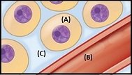
Additional info: The diagram visually distinguishes the three main fluid compartments and their spatial relationships.
