 Back
Foundations: An Introduction to Anatomy – Study Notes
Study Guide - Smart Notes
 Tailored notes based on your materials, expanded with key definitions, examples, and context.
Foundations: An Introduction to Anatomy
Definition and Scope of Anatomy
Anatomy is the scientific study of the external and internal structures of the body and the physical relationships among body parts. It involves careful observation and description of the human body, forming the foundation for understanding physiological processes and clinical practice.
External Anatomy: Study of structures visible on the outside of the body.
Internal Anatomy: Study of structures located within the body.
Physical Relationships: Understanding how body parts are connected and interact.
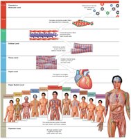
Levels of Organization in the Human Body
The human body is organized in a hierarchical manner, from the simplest chemical level to the most complex organismal level. Each level builds upon the previous one, allowing for increasing complexity and specialization.
Chemical or Molecular Level: Atoms and molecules essential for life (e.g., H, O, C, N make up 99% of human atoms).
Cellular Level: Smallest living units, composed of organelles that perform specific functions.
Tissue Level: Groups of similar cells working together to perform a specific function.
Organ Level: Structures composed of two or more tissue types working together (e.g., heart, liver).
Organ System Level: Groups of organs that perform related functions (e.g., digestive system).
Organism Level: The complete living human being.
The Eleven Organ Systems of the Human Body
Humans are composed of eleven distinct organ systems, each with specialized structures and functions. These systems work together to maintain homeostasis and overall health.
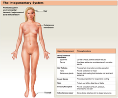
Integumentary System: Protects against environmental hazards and regulates body temperature.
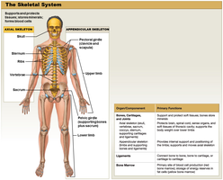
Skeletal System: Provides support, protection, and forms blood cells.
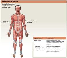
Muscular System: Allows movement, provides support, and produces heat.
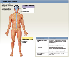
Nervous System: Directs immediate responses to stimuli and coordinates activities of other systems.
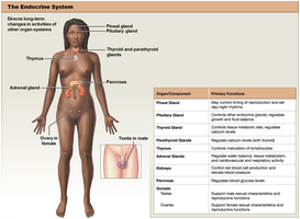
Endocrine System: Directs long-term changes in activities of other organ systems via hormones.
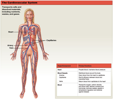
Cardiovascular System: Transports cells and dissolved materials, including nutrients, wastes, and gases.
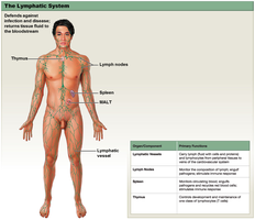
Lymphatic System: Defends against infection and disease, returns tissue fluid to bloodstream.
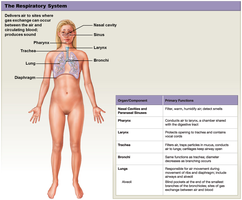
Respiratory System: Delivers air for gas exchange between air and circulating blood.
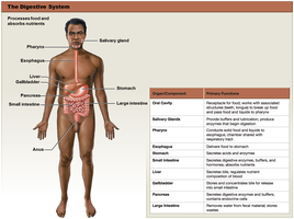
Digestive System: Processes food and absorbs nutrients.
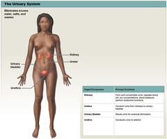
Urinary System: Eliminates excess water, salts, and wastes.
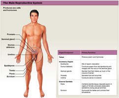
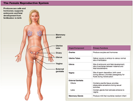
Reproductive System: Produces sex cells and hormones.
Importance of Precise Anatomical Terminology
Precise anatomical terminology is essential for clear communication among healthcare professionals. It ensures that descriptions of body locations, positions, and movements are universally understood, reducing errors in clinical and academic settings.
Anatomical Position and Body Orientation
The anatomical position is the standard reference for describing locations and directions on the human body. In this position, the individual stands upright, legs together, feet flat, arms at the sides, and palms facing forward with thumbs pointing outward.
Supine Position: Lying face up.
Prone Position: Lying face down.


Anatomical Landmarks
Specific terms are used to describe regions and landmarks on the body, aiding in precise localization of structures.
Cephalic: Head
Axillary: Armpit
Thoracic: Chest
Brachial: Arm
Antebrachial: Forearm
Cervical: Neck
Abdominal: Abdomen
Pelvic: Pelvis
Carpal: Wrist
Manual: Hand
Inguinal: Groin
Pubic: Genital region
Femoral: Thigh
Patellar: Kneecap
Crural: Leg (lower)
Tarsal: Ankle
Digital: Fingers or toes
Pedal: Foot
Lumbar: Lower back
Gluteal: Buttock
Sural: Calf
Plantar: Sole of foot
Abdominopelvic Quadrants and Regions
The abdominopelvic area is divided into four quadrants and nine regions to help locate underlying organs and describe pain or injury locations.
Four Quadrants: Right Upper, Left Upper, Right Lower, Left Lower
Nine Regions: Right/Left Hypochondriac, Epigastric, Right/Left Lumbar, Umbilical, Right/Left Inguinal, Hypogastric
Anatomical Directions
Anatomical directions are always described relative to the anatomical position and are paired as opposites for clarity.
Superior / Inferior: Above / Below
Lateral / Medial: Away from / Toward the midline
Proximal / Distal: Closer to / Farther from the point of attachment
Cranial (Cephalic) / Caudal: Toward the head / Toward the tail
Anterior (Ventral) / Posterior (Dorsal): Front / Back
Sectional Planes of the Body
Sectional planes are used to describe cuts or sections through the body, which are important for imaging and anatomical study.
Frontal (Coronal) Plane: Separates anterior and posterior portions.
Sagittal Plane: Separates right and left portions (midsagittal is exactly in the midline).
Transverse Plane: Separates superior and inferior portions (top and bottom).
Body Cavities
Body cavities protect internal organs and allow for changes in organ shape and size. Major cavities include:
Thoracic Cavity: Contains pleural cavities (lungs), mediastinum, and pericardial cavity (heart).
Abdominopelvic Cavity: Contains peritoneal, abdominal, and pelvic cavities.
Serous Membranes: Visceral and Parietal Layers
Many body cavities are lined with serous membranes, which have two layers:
Visceral Layer: Inner membrane that touches the organ.
Parietal Layer: Outer membrane that lines the cavity wall.
Serous Fluid: Lubricates and reduces friction between the two layers.
