 Back
BackFoundations and Modern Concepts in Anatomy and Physiology: From Historical Roots to Cellular Structure
Study Guide - Smart Notes
 Tailored notes based on your materials, expanded with key definitions, examples, and context.
A Brief History of Anatomy
Early Contributions and the House of Wisdom
The study of anatomy has ancient roots, with significant advancements made during the Islamic Golden Age at the House of Wisdom. Scholars here translated and critiqued classical texts, laying the groundwork for modern science.
al-Khwarizmi: Authored 'Kitab al-Jebr,' foundational for number theory.
al-Kindi: Introduced the Hindu decimal system to the Islamic world.
Ibn Sina (Avicenna): Wrote 'The Canon of Medicine,' critically evaluating earlier medical authorities like Galen.

Renaissance and the Rebirth of Anatomy
The Renaissance (1400–1600) marked a revival of scientific inquiry in Europe, fueled by the rediscovery of ancient texts and a spirit of experimentation.
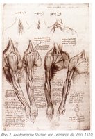
Leonardo da Vinci: Produced the first detailed anatomical drawings based on dissections, including studies of motion.
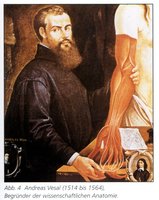
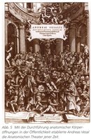
Andreas Vesalius: Revolutionized anatomy with public dissections and his work 'De fabrica humani corporis.'
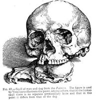
Hieronymus Fabricius: Compared human and animal anatomy, such as skull structures.
The Early Scientific Era (1600–1800): Experimentation
This era emphasized experimentation and observation, leading to foundational discoveries in physiology and microscopy.
William Harvey: Demonstrated the circulation of blood, overturning previous models of blood flow.
Antony van Leeuwenhoek: Developed the first usable microscope, discovering microbial life.
Robert Hooke: Invented the compound microscope and coined the term 'cell.'
Carl Zeiss: Improved microscopes, enabling detailed histological studies.

Evolutionary Anatomy in the 19th Century
The 19th century saw the integration of evolutionary theory into anatomy, greatly expanding the field's scope.
Charles Darwin: Proposed the theory of evolution by natural selection.
Huxley: Advanced comparative anatomy.
Karl Ernst von Baer: Studied embryological development.
Gregor Mendel: Established the principles of heredity.
T.H. Morgan: Developed the modern concept of genetics.
Modern and 21st Century Anatomy
Contemporary anatomy explores how genetic and environmental factors shape development, with a focus on personalized medicine and advanced imaging techniques.
Key questions include: How do genes direct anatomical development? How does the environment influence structure?
The Cell: The Living Unit
Objectives
Understanding the cell is fundamental to anatomy and physiology. Key objectives include:
Familiarity with cell theory
Knowledge of cellular components and their functions
Recognition of active and passive transport mechanisms
Cell Theory
Formulated by Matthias Schleiden and Theodor Schwann, cell theory is a cornerstone of biology:
The cell is the basic unit of structure, physiology, and organization in living things.
Cells retain a dual existence as distinct entities and as building blocks of organisms.
All cells arise from pre-existing cells.
All metabolic events occur within cells.
Additional info: This theory underpins modern biomedical research and clinical practice.
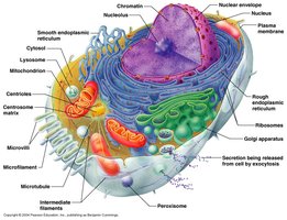
The (Not So) 'Typical' Cell
Cells vary greatly in structure and function, but share common features such as a plasma membrane, cytoplasm, and organelles.
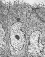
Transmission Electron Micrograph of a Eukaryotic Cell
Electron microscopy reveals the complex internal organization of eukaryotic cells, including distinct apical and basolateral domains.
Apical domain: Involved in protection, absorption, and secretion.
Basolateral domain: Provides anchorage and communication with neighboring cells.
Cytoplasm
The cytoplasm is the material between the plasma membrane and the nucleus, consisting of:
Cytosol: Aqueous solution with dissolved proteins, salts, sugars, and other solutes.
Cytoplasmic organelles: Specialized structures for metabolic processes.
Inclusions: Stored nutrients and pigments.
The hydrostatic properties of the cytoplasm help maintain cell shape, and its consistency varies with cell function.
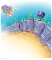
The Plasma Membrane
Structure and Function
The plasma membrane is a dynamic, selectively permeable barrier that separates the cell's interior from its external environment.
Composed of a lipid bilayer with embedded proteins (fluid mosaic model).
Separates cytoplasm (intracellular fluid) from interstitial (extracellular) fluid.
Plays a key role in cellular communication, transport, and signaling.

Membrane Lipids
Phospholipids (75%): Form the bilayer; phosphate heads are polar and hydrophilic, fatty acid tails are nonpolar and hydrophobic.
Glycolipids (5%): Lipids with attached sugar groups, found on the outer surface.
Cholesterol (20%): Stabilizes membrane fluidity and integrity.
Membrane Proteins
Integral proteins: Firmly embedded, often span the membrane; function as channels, carriers, enzymes, or receptors.
Peripheral proteins: Loosely attached to integral proteins; function as enzymes, structural anchors, or in cell signaling.
Functions of Membrane Proteins
Transport: Move substances across the membrane.
Receptors for signal transduction: Bind signaling molecules and initiate cellular responses.
Attachment to cytoskeleton and extracellular matrix: Maintain cell shape and stabilize location.
Enzymatic activity: Catalyze specific reactions at the membrane surface.
Intercellular joining: Facilitate cell-cell adhesion.
Cell-cell recognition: Allow cells to identify each other.
