 Back
Foundations of Anatomy: Body Organization, Cells, and Tissues
Study Guide - Smart Notes
 Tailored notes based on your materials, expanded with key definitions, examples, and context.
Foundations: An Introduction to Anatomy
Overview of Anatomy and Physiology
Anatomy is the study of the structure of the body, both externally and internally, while physiology focuses on how these structures function. Understanding both is essential for comprehending how the human body operates and adapts throughout life.
Gross Anatomy: Study of structures visible without magnification, including surface and regional anatomy.
Microscopic Anatomy: Study of structures requiring magnification, such as cytology (cells) and histology (tissues).
Developmental Anatomy: Examines changes from embryo to adult, including embryology and specialization of anatomical structures.
Comparative Anatomy: Considers similarities and differences between species.
Clinical Anatomy: Studies anatomical changes during illness.
Surgical Anatomy: Focuses on landmarks important for surgery.
Radiographic and Cross-sectional Anatomy: Uses imaging techniques (e.g., CT, MRI) to study anatomy.
Levels of Organization
The human body is organized into hierarchical levels, each building upon the previous.
Chemical/Molecular Level: Atoms and molecules form the basis of cellular structures.
Cellular Level: Cells are the smallest functional units.
Tissue Level: Groups of similar cells performing specific functions.
Organ Level: Combination of tissues forming organs.
Organ System Level: Multiple organs working together (e.g., digestive system).
Features of Living Organisms: Responsiveness, growth, differentiation, reproduction, movement, metabolism, excretion, absorption, respiration.
Overview of Organ Systems
The body contains eleven major organ systems, each with distinct functions:
Integumentary System: Protection against environmental hazards.
Skeletal System: Support, protection, and movement.
Muscular System: Locomotion and heat production.
Nervous System: Directs responses to stimuli.
Cardiovascular System: Transports cells and materials.
Respiratory System: Delivers air for gas exchange.
Digestive System: Processes food.
Urinary System: Eliminates waste.
Reproductive System: Produces sex cells and hormones.
Directional Terms and Anatomical Regions
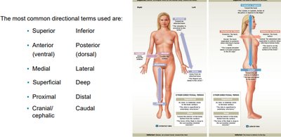
Directional terms are used to describe locations and relationships of body parts.
Superior/Inferior: Above/below.
Anterior (ventral)/Posterior (dorsal): Front/back.
Medial/Lateral: Toward midline/away from midline.
Superficial/Deep: Near surface/away from surface.
Proximal/Distal: Closer to/farther from point of attachment.
Cranial/Caudal: Toward head/toward tail.
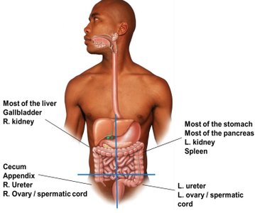
The abdominopelvic region is divided into quadrants and nine regions for clinical reference.
Quadrants: Right/Left, Upper/Lower.
Regions: Right/Left hypochondriac, epigastric, right/left lumbar, umbilical, right/left iliac, hypogastric.
Planes and Sections
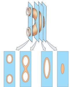
Body planes are used to describe anatomical sections:
Sagittal Plane: Divides body into left and right.
Midsagittal: Equal left and right halves.
Parasagittal: Unequal left and right.
Transverse Plane: Divides body into superior and inferior.
Frontal Plane: Divides body into anterior and posterior.
Serial Reconstruction: Series of sections at small intervals.
Body Cavities
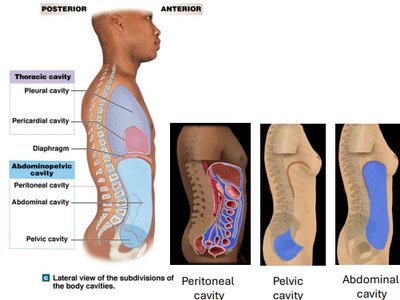
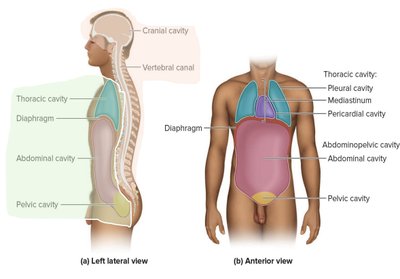
The body contains two major cavities: posterior (dorsal) and anterior (ventral).
Posterior (Dorsal): Contains cranial and spinal cavities.
Anterior (Ventral): Contains thoracic and abdominopelvic cavities, separated by the diaphragm.
Thoracic Cavity: Heart, lungs, pleural, pericardial, and mediastinum.
Abdominopelvic Cavity: Abdominal, pelvic, and peritoneal cavities.
Foundations: The Cell
Cell Structure and Function
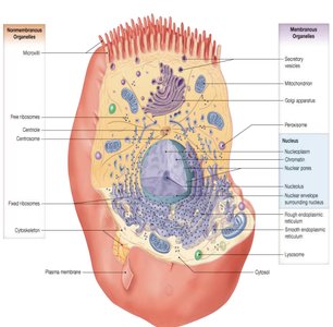
Cells are the basic building blocks of all living organisms. They are classified as sex cells or somatic cells and consist of cytoplasm and a plasma membrane.
Cytosol: Fluid inside the cell.
Organelles: Intracellular structures with specific functions.
Plasma Membrane: Composed of phospholipids, glycolipids, sterols, and proteins.

Plasma Membrane
The plasma membrane provides a physical barrier, regulates material exchange, responds to changes in extracellular fluid, and supports cell communication and adhesion.
Phospholipid Bilayer: Hydrophilic heads face outward, hydrophobic tails inward.
Proteins: Peripheral (attached to membrane surface) and integral (embedded, forming channels).
Glycolipids and Sterols: Form glycocalyx coating.
Membrane Permeability
Membrane permeability can be passive or active:
Passive: Diffusion, facilitated diffusion, osmosis.
Active: Active transport, endocytosis, exocytosis (require ATP).
Foundations: Tissues and Early Embryology
Epithelial Tissue
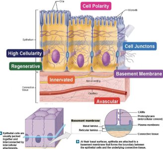
Epithelial tissue consists of closely bound cells with exposed apical surfaces and attached basal surfaces. It is avascular, provides protection, controls permeability, and produces secretions.
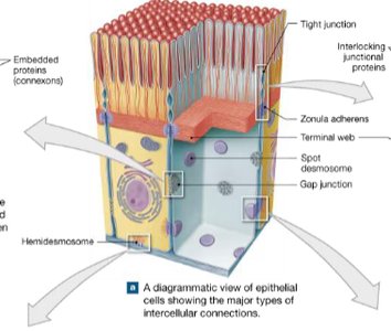
Intercellular Connections: Tight junctions, desmosomes, gap junctions.
Attachment to Basal Lamina: Maintains tissue integrity.
Regeneration: Frequent replacement via stem cells.
Classification of Epithelia
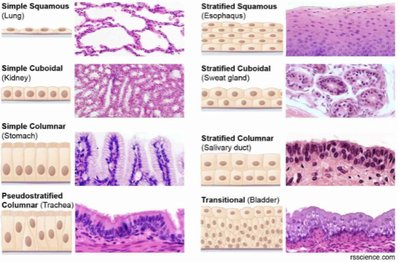
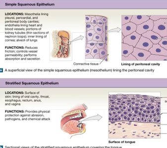
Epithelia are classified by cell shape and layering:
Simple: One layer (e.g., lungs, kidney).
Stratified: Multiple layers (e.g., esophagus, sweat gland).
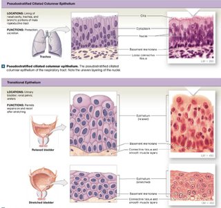
Pseudostratified: Appears layered, nuclei at different levels (e.g., trachea).
Transitional: Many layers, can stretch (e.g., bladder).
Glandular Epithelia
Glandular epithelia produce secretions and are classified as unicellular (goblet, mucous cells) or multicellular glands. Modes of secretion include eccrine, apocrine, and holocrine.
Connective Tissue
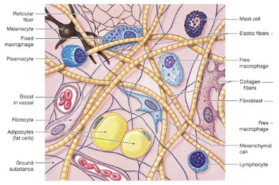
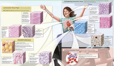
Connective tissue supports, protects, and binds other tissues. It is divided into:
Connective Tissue Proper: Loose (open framework) and dense (packed elastic).
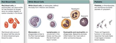
Fluid Connective Tissue: Blood and lymph.
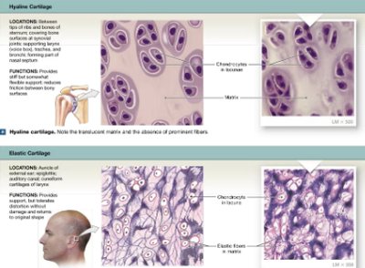
Supporting Connective Tissue: Cartilage and bone.
Nervous Tissue
Nervous tissue conducts electrical impulses and consists of neurons (impulse transmission) and neuroglia (support and nutrition).
Muscle Tissue
Muscle tissue facilitates movement and includes skeletal, cardiac, and smooth muscle types.
Membranes
Membranes are formed by epithelial and connective tissues and include mucous, serous, cutaneous, and synovial membranes.
Stem Cells and Early Embryology
Stem cells are classified as embryonic (totipotent, pluripotent) or adult (multipotent, unipotent). Fertilization leads to blastocyst formation and implantation, followed by gastrulation and formation of three germ layers: ectoderm, mesoderm, endoderm.
The Integumentary System
Structure and Development
The integumentary system includes the skin, hair, nails, and glands. It consists of four tissue types: epithelium, connective, smooth muscle, and nervous tissue. The skin develops from ectoderm and mesoderm, forming epidermis, dermis, and accessory structures.
Epidermis
The epidermis is divided into five layers: stratum basale, stratum spinosum, stratum granulosum, stratum lucidum (thick skin only), and stratum corneum. Skin color is determined by carotene and melanin, and dermal ridges form fingerprints.
Dermis and Hypodermis
The dermis contains papillary and reticular layers, providing strength, elasticity, and blood supply. The hypodermis stabilizes the skin and contains adipose tissue.
Accessory Structures
Accessory structures include hair follicles, glands (sebaceous, sweat), and nails. Hair provides protection and sensory input, while glands assist in lubrication and thermoregulation.
The Skeletal System: Osseous Tissue and Bone Structure
Functions and Structure
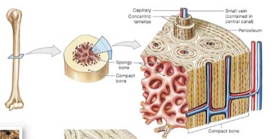
The skeletal system provides support, protection, movement, mineral storage, and blood cell production. Bones are dynamic organs composed of osseous tissue, cartilage, ligaments, and connective tissue.
Bone Matrix and Cells
The bone matrix is composed of calcium phosphate (hydroxyapatite crystals) and collagen fibers. Bone cells include osteocytes (mature), osteoblasts (immature, bone formation), osteoprogenitor cells (stem cells), and osteoclasts (bone resorption).
Bone Growth and Development
Bones develop through intramembranous and endochondral ossification. Growth occurs via appositional growth and is regulated by minerals, vitamins, and hormones.
Accessory Structures in Joints
Joints are supported by cartilage, ligaments, tendons, menisci, fat pads, and bursae. Types of cartilage include hyaline and fibrocartilage, each with specific locations and functions.
The Muscular System: Skeletal Muscle Tissue and Muscle Organization
Properties of Muscle Tissue
Muscle tissue exhibits excitability, contractility, extensibility, and elasticity, enabling movement and force generation.
*Additional info: Academic context and explanations have been expanded for clarity and completeness. Only images directly relevant to the adjacent content have been included as per strict relevance guidelines.*
