 Back
BackFoundations of Human Anatomy: Introduction, Terminology, and Body Structure
Study Guide - Smart Notes
 Tailored notes based on your materials, expanded with key definitions, examples, and context.
Introduction to Anatomy
Definition and Scope
Anatomy is the scientific discipline concerned with the organization and structure of the human body. The term originates from Greek, meaning "to cut up," reflecting its historical roots in dissection. Modern anatomy encompasses both macroscopic (gross) and microscopic structures, utilizing advanced technologies such as electron microscopes, CT scans, and MRIs.
Macroscopic (Gross) Anatomy: Study of structures visible to the naked eye.
Microscopic Anatomy: Study of structures requiring magnification, including cytology (cells) and histology (tissues).
Functional Significance: Anatomy provides the foundation for understanding physiology—the function of body structures.
Divisions of Anatomy
Surface Anatomy: General form and superficial markings.
Systemic Anatomy: Study of body systems (e.g., digestive, nervous).
Regional Anatomy: Study of specific regions and all tissues within them.
Developmental Anatomy: Structural changes from fertilized egg to adult; includes embryology.
Medical/Radiological Anatomy: Changes during disease and features visible via radiographic technology.
Generalized Body Structure
Body Divisions
The human body is organized into hollow spaces (cavities) and solid structures. The main divisions are:
Body Wall: Framework supporting and enclosing organs (skin, skeleton, muscles).
Body Cavities: Internal spaces housing organs; largest is the ventral cavity (thoracic and abdominopelvic).
Organs: Structures capable of specific functions, located within cavities or as part of the body wall.
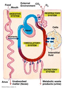
Body Systems
Organs with similar functions are grouped into body systems. There are eleven major systems:
Integumentary System: Skin, hair, nails; protection and temperature control.
Skeletal System: Bones, ligaments, cartilage; support, protection, blood formation.
Muscular System: Skeletal muscles, tendons; movement and heat production.
Nervous System: Brain, spinal cord, nerves; control and perception.
Endocrine System: Glands; chemical coordination via hormones.
Cardiovascular System: Heart, blood vessels; transport of nutrients, gases, waste.
Lymphatic System: Lymph vessels, nodes; defense and blood volume maintenance.
Respiratory System: Lungs, airways; gas exchange.
Digestive System: GI tract, organs; food processing and nutrient absorption.
Urinary System: Kidneys, bladder; regulation of blood chemistry and waste elimination.
Reproductive System: Sex organs; production and support of sex cells and hormones.
Anatomical Terminology
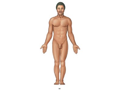
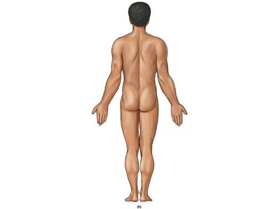
Standard Anatomical Position (SAP)
All anatomical descriptions assume the Standard Anatomical Position:
Standing erect
Upper limbs at sides
Lower limbs together
Face, palms, and feet directed forward

Principle Body Regions
Anatomical terms describe specific regions and areas of the body. Knowing both anatomical and common names is essential for clear communication.
Cephalon (Head): Cranium (skull), Nasus (nose), Bucca (cheek), Auris (ear), Oculus (eye), Oris (mouth)
Cervicis (Neck)
Thoracis (Chest): Mamma (breast), Axilla (armpit), Brachium (arm), Antebrachium (forearm), Carpus (wrist), Manus (hand), Palma (palm), Digits (fingers)
Abdomen (Abdominal region)
Pelvis (Pelvic region): Coxa (hip), Pubis (anterior pelvis), Inguen (groin)
Lumbus (Lower back), Gluteus (buttock), Femur (thigh), Patella (knee), Popliteus (back of knee), Crus (leg), Sura (calf), Tarsus (ankle), Pes (foot), Planta (sole), Digits (toes), Calcaneus (heel)
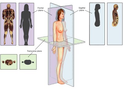
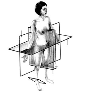
Planes of Section
Body Planes
The body can be divided along imaginary planes for anatomical study:
Sagittal Plane: Divides right and left sections
Midsagittal Plane: Equal right and left halves
Parasagittal Plane: Unequal right and left sections
Frontal (Coronal) Plane: Anterior and posterior sections
Transverse (Horizontal) Plane: Superior and inferior sections
Oblique Plane: Angled sections
Directional Terms
Relative Location
Directional terms describe the relationship between body parts:
Superior: Above; toward the head
Inferior: Below; toward the feet
Cephalic/Cranial: Toward the head
Anterior (Ventral): Near the front
Posterior (Dorsal): Near the back
Medial: Toward the midline
Lateral: Away from the midline
Ipsilateral: Same side
Contralateral: Opposite side
Proximal: Near attachment point
Distal: Far from attachment point
Superficial: Toward the surface
Intermediate: Between superficial and deep
Deep: Away from the surface
Body Cavities
Major Cavities and Their Functions
Body cavities house, protect, and allow movement of organs. They also enable different internal pressures.
Dorsal Body Cavity: Cushions and protects the central nervous system
Cranial Cavity: Houses the brain
Spinal (Vertebral) Cavity: Houses the spinal cord
Ventral Body Cavity (Coelom): Enclosed by ribs and muscles; surrounds organs of respiratory, digestive, cardiovascular, urinary, and reproductive systems
Thoracic Cavity: Houses heart and lungs
Abdominopelvic Cavity: Houses abdominal and pelvic organs
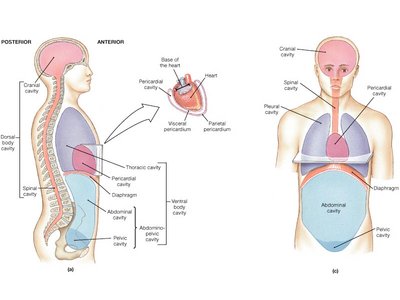
Serous Membranes
Structure and Function
Serous membranes line all body cavities and organs, secreting serous fluid for frictionless movement. They are named by location:
Visceral Serous Membranes: Cover organs (e.g., visceral pleura for lungs, visceral pericardium for heart, visceral peritoneum for abdominal organs)
Parietal Serous Membranes: Line cavities (e.g., parietal pleura for pleural cavity, parietal pericardium for pericardial cavity, parietal peritoneum for abdominopelvic cavity)
Mediastinum: Space between pleural cavities, containing the pericardial cavity and other structures
Clinical Significance
Peritonitis: Inflammation of the abdominal peritoneum, often due to injury or infection, can cause fluid accumulation (ascites) and symptoms such as heartburn, indigestion, and lower back pain.
Key Definitions
Anatomy: Study of body structure
Histology: Study of tissues
Body Cavity: Internal chamber housing organs
Standard Anatomical Position: Reference posture for anatomical descriptions
Midsagittal Plane: Divides body into equal right and left halves
Serous Membrane: Fluid-secreting membrane lining cavities and organs
Mediastinum: Central thoracic space between pleural cavities
Summary Table: Body Systems and Functions
System | Main Components | Function |
|---|---|---|
Integumentary | Skin, hair, nails | Protection, temperature control |
Skeletal | Bones, ligaments | Support, protection, blood formation |
Muscular | Muscles, tendons | Movement, heat production |
Nervous | Brain, spinal cord, nerves | Control, perception |
Endocrine | Glands | Coordination via hormones |
Cardiovascular | Heart, blood vessels | Transport of materials |
Lymphatic | Lymph vessels, nodes | Defense, blood volume |
Respiratory | Lungs, airways | Gas exchange |
Digestive | GI tract, organs | Food processing, absorption |
Urinary | Kidneys, bladder | Waste elimination, blood chemistry |
Reproductive | Sex organs | Production of sex cells, hormones |
Summary Table: Anatomical Planes
Plane | Description |
|---|---|
Sagittal | Right and left sections |
Midsagittal | Equal right and left halves |
Parasagittal | Unequal right and left sections |
Frontal (Coronal) | Anterior and posterior sections |
Transverse | Superior and inferior sections |
Oblique | Angled sections |
Summary Table: Directional Terms
Term | Definition |
|---|---|
Superior | Above; toward the head |
Inferior | Below; toward the feet |
Anterior (Ventral) | Near the front |
Posterior (Dorsal) | Near the back |
Medial | Toward the midline |
Lateral | Away from the midline |
Proximal | Near attachment point |
Distal | Far from attachment point |
Superficial | Toward the surface |
Deep | Away from the surface |
Summary Table: Body Cavities
Cavity | Location | Contents |
|---|---|---|
Cranial | Skull | Brain |
Spinal | Vertebral column | Spinal cord |
Thoracic | Chest | Heart, lungs |
Abdominal | Abdomen | Digestive organs |
Pelvic | Pelvis | Urinary, reproductive organs |
Additional info: Academic context was added to clarify the functional significance of anatomical divisions, body systems, and serous membranes, as well as to provide self-contained explanations for exam preparation.
