 Back
Functional Anatomy of Endocrine Glands: Structure, Hormones, and Histology
Study Guide - Smart Notes
 Tailored notes based on your materials, expanded with key definitions, examples, and context.
Introduction to the Endocrine System
The endocrine system is a network of glands that secrete hormones directly into the bloodstream to regulate various physiological processes. These hormones control growth, metabolism, reproduction, and homeostasis. Understanding the structure and function of each gland is essential for comprehending how the body maintains internal balance.
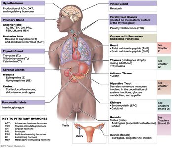
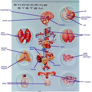
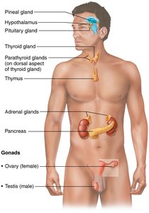
Major Endocrine Glands: Hypothalamus, Pituitary gland, Thyroid gland, Parathyroid glands, Adrenal glands, Pancreas, Ovaries, Testes
Key Concepts: Identification of glands, hormones produced, major functions, and target organs/cells
Pituitary Gland (Hypophysis)
Structure and Relationship to Hypothalamus
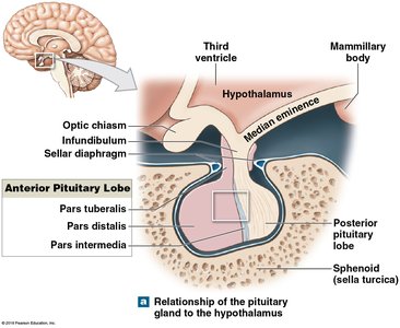
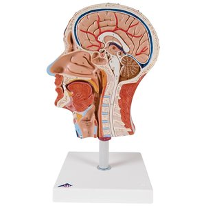
The pituitary gland, often called the "master gland," is located at the base of the brain and is connected to the hypothalamus. It consists of two main lobes: the anterior pituitary (adenohypophysis) and the posterior pituitary (neurohypophysis). The hypothalamus regulates pituitary function through releasing and inhibiting hormones.
Anterior Pituitary (Adenohypophysis): Produces tropic and general hormones
Posterior Pituitary (Neurohypophysis): Stores and releases hormones produced by the hypothalamus
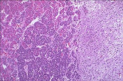
Histological Features
Anterior Pituitary: Appears more cellular and granular due to hormone-producing cells
Posterior Pituitary: Contains more nerve fibers and appears lighter
Pituitary Hormones, Targets, and Effects
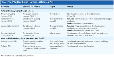
The pituitary gland secretes several hormones, each with specific targets and physiological effects. Tropic hormones primarily regulate other endocrine glands, while general hormones affect various tissues.
Hormone | Stimulus for Release | Target | Effects |
|---|---|---|---|
Thyroid-stimulating hormone (TSH) | Thyrotropin-releasing hormone (TRH) | Thyroid gland | Stimulates secretion of thyroid hormones |
Follicle-stimulating hormone (FSH) | Gonadotropin-releasing hormone (GnRH) | Ovaries and testes | Females: ovarian follicle maturation and estrogen; Males: sperm production |
Luteinizing hormone (LH) | Gonadotropin-releasing hormone (GnRH) | Ovaries and testes | Females: ovulation and estrogen/progesterone; Males: testosterone production |
Adrenocorticotropic hormone (ACTH) | Corticotropin-releasing hormone (CRH) | Adrenal cortex | Stimulates release of glucocorticoids and androgens |
Growth hormone (GH) | Growth hormone-releasing hormone (GHRH) | Liver, muscle, bone, cartilage | Stimulates growth and protein synthesis |
Prolactin (PRL) | A decrease in prolactin-inhibiting hormone | Mammary glands | Stimulates milk production |
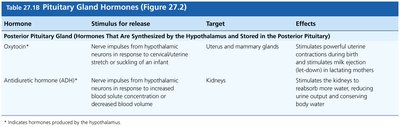
Hormone | Stimulus for Release | Target | Effects |
|---|---|---|---|
Oxytocin (OXT) | Nerve impulses from hypothalamus | Uterus, mammary glands | Stimulates uterine contractions and milk ejection |
Antidiuretic hormone (ADH) | Nerve impulses from hypothalamus | Kidneys | Stimulates water reabsorption, reduces urine output |
Thyroid and Parathyroid Glands
Structure and Identification
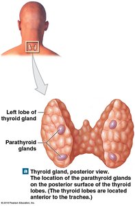
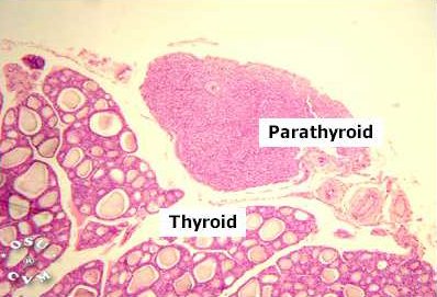
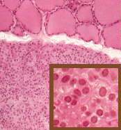
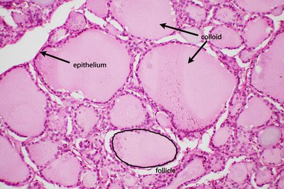
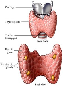
The thyroid gland is located in the anterior neck, consisting of two lobes connected by an isthmus. The parathyroid glands are small, oval glands embedded on the posterior surface of the thyroid. Histologically, the thyroid contains spherical follicles lined by follicular cells, while the parathyroid contains principal (chief) cells.
Thyroid Follicles: Spheres lined by simple cuboidal epithelium (follicular cells) that produce thyroid hormones (T3 and T4)
Parathyroid Glands: Principal cells produce parathyroid hormone (PTH)
Hormones, Targets, and Effects
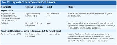
Hormone(s) | Stimulus for Release | Target | Effects |
|---|---|---|---|
Thyroxine (T4), Triiodothyronine (T3) | Thyroid-stimulating hormone (TSH) | Most cells of the body | Increase basal metabolic rate (BMR), regulate growth and development |
Calcitonin | High blood calcium | Bones | Lowers blood calcium (pharmacological effect) |
Parathyroid hormone (PTH) | Low blood calcium | Bones, kidneys | Increases blood calcium by stimulating osteoclasts and kidney reabsorption |
Adrenal Glands
Structure and Histology
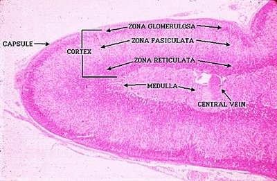
The adrenal glands are located atop the kidneys and consist of an outer cortex and an inner medulla. The cortex is divided into three zones, each producing different hormones, while the medulla produces catecholamines.
Zona Glomerulosa (outer): Produces mineralocorticoids (e.g., aldosterone)
Zona Fasciculata (middle): Produces glucocorticoids (e.g., cortisol, cortisone)
Zona Reticularis (inner): Produces androgens
Medulla: Produces epinephrine and norepinephrine
Pancreas
Structure and Endocrine Function
The pancreas is a mixed gland with both exocrine and endocrine functions. The endocrine portion consists of clusters called pancreatic islets (Islets of Langerhans), which contain alpha and beta cells.
Alpha (α) cells: Produce glucagon, which raises blood glucose
Beta (β) cells: Produce insulin, which lowers blood glucose
Note: Only the endocrine function is covered here; exocrine function is discussed in the digestive system chapter.
Gonads: Testes and Ovaries
Testes
Produce sperm and androgens (e.g., testosterone)
Ovaries
Produce eggs, estrogens, and progesterone
Note: Gonads are covered in detail in the reproductive system chapter; here, focus is on their endocrine function.
Sample Lab Practical Questions
Which portion of the pituitary gland is highlighted? Answer: Posterior pituitary
Which structures are highlighted? Answer: Thyroid follicles
Which of the following hormones is produced from the highlighted organ? Answer: Glucagon
What is the result of secretions from the highlighted region? Answer: Increase blood pressure and heart rate
