 Back
Functional Anatomy of the Brain and Cranial Nerves: Structured Study Notes
Study Guide - Smart Notes
 Tailored notes based on your materials, expanded with key definitions, examples, and context.
The Brain, Cranial Nerves, and Sensory and Motor Pathways
Origins and Development of Brain Regions
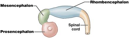
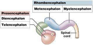
The brain develops from the embryonic neural tube, which forms primary and secondary brain vesicles. These vesicles give rise to the major regions of the adult brain, each with distinct structural and functional characteristics.
Primary Brain Vesicles: Prosencephalon (forebrain), Mesencephalon (midbrain), Rhombencephalon (hindbrain).
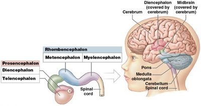
Secondary Brain Vesicles: Prosencephalon subdivides into the diencephalon and telencephalon; Rhombencephalon subdivides into the metencephalon and myelencephalon.
Largest Region: The telencephalon develops into the cerebrum, the largest part of the adult brain.
Major Regions of the Brain
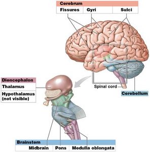
The adult brain is divided into four major regions, each with specialized functions:
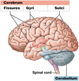
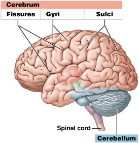
Cerebrum: Responsible for conscious thought, memory, sensory processing, and control of skeletal muscles. Characterized by gyri (folds) and sulci (grooves) that increase surface area.
Cerebellum: Coordinates and modulates motor commands, maintaining balance and fine-tuning movements.
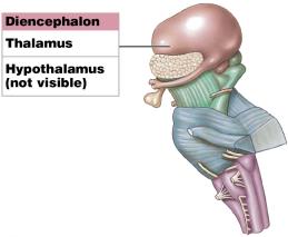
Diencephalon: Includes the thalamus (sensory relay/processing) and hypothalamus (emotions, autonomic function, hormone production).
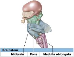
Brainstem: Composed of the midbrain, pons, and medulla oblongata. Controls reflexes, relays information, and regulates vital autonomic functions.
Ventricular System of the Brain
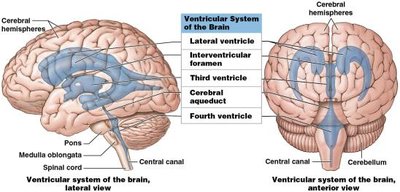
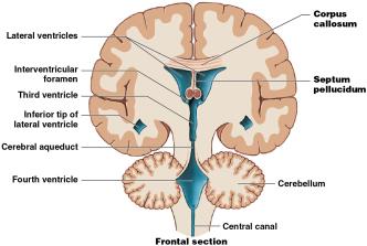
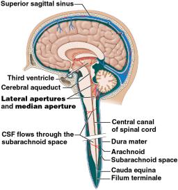
The brain contains interconnected chambers called ventricles, which are filled with cerebrospinal fluid (CSF) and lined by ependymal cells. The ventricular system supports and protects the brain.
Lateral Ventricles: Located in each cerebral hemisphere.
Third Ventricle: Located in the diencephalon.
Cerebral Aqueduct: Connects the third and fourth ventricles.
Fourth Ventricle: Extends from the metencephalon to the medulla oblongata; narrows to become the central canal of the spinal cord.
Interconnections: Corpus callosum (white matter tract) interconnects hemispheres; septum pellucidum separates lateral ventricles.
Protection and Support of the Brain
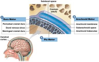
The brain is protected by cranial bones, cerebrospinal fluid, the blood-brain barrier, and cranial meninges. The meninges are three layers that surround the brain and spinal cord:
Dura Mater: Two fibrous layers; periosteal layer fused to cranial bones, meningeal layer forms dural folds and venous sinuses.
Arachnoid Mater: Membrane closest to dura mater; contains subarachnoid space and trabeculae.
Pia Mater: Bound to the brain surface by astrocyte processes; extends into every fold and accompanies blood vessels.
Dural Folds and Venous Sinuses
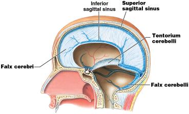
Falx Cerebri: Separates cerebral hemispheres; contains superior and inferior sagittal sinuses.
Tentorium Cerebelli: Separates cerebrum from cerebellum.
Falx Cerebelli: Separates cerebellar hemispheres.
Dural Venous Sinuses: Large veins within dural folds; superior sagittal sinus is the largest.
Cerebrospinal Fluid (CSF)
Functions: Supports brain weight, cushions from trauma, transports nutrients, chemical messengers, and wastes.
Production: Produced and maintained by choroid plexus (ependymal cells and capillaries) in each ventricle.
Circulation: CSF circulates from choroid plexus through ventricles and central canal, then is absorbed into venous circulation at arachnoid granulations.
Summary Table: Major Brain Regions and Functions
Region | Main Function |
|---|---|
Cerebrum | Conscious thought, memory, sensory processing, motor control |
Cerebellum | Coordination and modulation of motor commands |
Diencephalon | Sensory relay (thalamus), autonomic and endocrine control (hypothalamus) |
Brainstem | Reflexes, relay of information, regulation of vital functions |
Additional info:
The notes above expand on brief lecture points to provide full academic context, including definitions, examples, and structural-functional relationships.
Images included are directly relevant to the anatomical structures and processes described, reinforcing visual understanding of brain development, regions, protection, and ventricular system.
