 Back
BackFunctional Anatomy of the Brain and Nervous System
Study Guide - Smart Notes
 Tailored notes based on your materials, expanded with key definitions, examples, and context.
Central Nervous System (CNS)
Overview of Brain Regions
The central nervous system (CNS) is composed of the brain and spinal cord. The brain is divided into several major regions, each with specialized functions essential for sensory processing, motor control, and higher cognitive functions.
Cerebral hemispheres
Diencephalon
Brain stem
Cerebellum
Cerebral Hemispheres
Structure and Surface Features
The cerebral hemispheres are the paired superior parts of the brain, accounting for more than half of its mass. The surface is characterized by ridges and grooves that increase the surface area for neural processing.
Gyri: Elevated ridges on the brain surface.
Sulci: Shallow grooves between gyri.
Fissures: Deeper grooves that separate large regions of the brain.
Lobes: Named for the cranial bones overlying them (frontal, parietal, temporal, occipital).
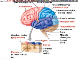
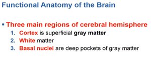
Main Regions of the Cerebral Hemisphere
The cerebral hemispheres are organized into three main regions, each with distinct functions and structures.
Cortex: Superficial gray matter responsible for processing sensory information and higher cognitive functions.
White matter: Deeper tissue composed of myelinated nerve fibers that connect different brain regions.
Basal nuclei: Deep pockets of gray matter involved in motor control.
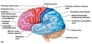
Functional Areas of the Cerebral Cortex
The cerebral cortex contains specialized areas responsible for sensory perception, voluntary movement, and complex cognitive tasks.
Primary motor area: Controls voluntary movements.
Primary somatic sensory area: Receives and processes sensory input from the body.
Association areas: Involved in higher functions such as memory, judgment, and language.
Special sensory areas: Visual, auditory, gustatory, and olfactory processing.
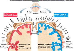
Motor and Sensory Maps
The precentral gyrus (motor cortex) and postcentral gyrus (sensory cortex) contain maps representing different body regions. The size of each region reflects the degree of motor control or sensory sensitivity.
Motor map: Located in the precentral gyrus, controls voluntary movements.
Sensory map: Located in the postcentral gyrus, processes sensory input.
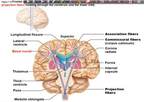
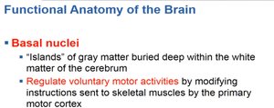
White Matter and Basal Nuclei
White matter consists of fiber tracts that connect different brain regions. Basal nuclei are clusters of gray matter deep within the white matter, playing a key role in regulating voluntary motor activities.
Association fibers: Connect areas within the same hemisphere.
Commissural fibers: Connect the two hemispheres (e.g., corpus callosum).
Projection fibers: Connect the cerebrum with lower CNS regions.
Basal nuclei: Modify instructions sent to skeletal muscles by the primary motor cortex.
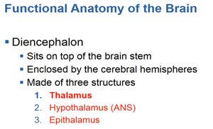
Diencephalon
Overview and Main Structures
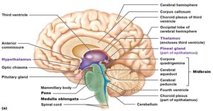
The diencephalon sits atop the brain stem and is enclosed by the cerebral hemispheres. It is composed of three main structures: the thalamus, hypothalamus, and epithalamus.
Thalamus
Hypothalamus
Epithalamus
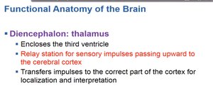
Thalamus
The thalamus encloses the third ventricle and acts as a relay station for sensory impulses ascending to the cerebral cortex. It directs impulses to the appropriate cortical areas for localization and interpretation.
Relay station for sensory information
Transfers impulses for localization and interpretation
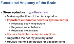
Hypothalamus
The hypothalamus forms the floor of the diencephalon and is a major autonomic nervous system center. It regulates body temperature, water balance, metabolism, and houses the limbic center for emotions. It also controls the pituitary gland and mammillary bodies for olfaction.
Regulates body temperature, water balance, metabolism
Controls the pituitary gland
Houses the limbic center for emotions
Involved in olfaction (smell)
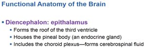
Epithalamus
The epithalamus forms the roof of the third ventricle, houses the pineal body (an endocrine gland), and includes the choroid plexus, which forms cerebrospinal fluid.
Pineal body: Secretes melatonin, regulates sleep-wake cycles
Choroid plexus: Produces cerebrospinal fluid
Brain Stem
Overview and Main Parts
The brain stem connects the brain to the spinal cord and is composed of the midbrain, pons, and medulla oblongata. It is responsible for basic life-sustaining functions.
Midbrain
Pons
Medulla oblongata
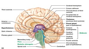
Midbrain
The midbrain extends from the mammillary bodies to the pons and contains the cerebral aqueduct. It includes fiber tracts for impulse conduction and the corpora quadrigemina, which are visual and auditory reflex centers.
Cerebral aqueduct: Connects third and fourth ventricles
Cerebral peduncles: Convey impulses
Corpora quadrigemina: Visual and auditory reflex centers
Pons
The pons is a rounded structure below the midbrain, composed mainly of fiber tracts. It contains nuclei involved in the control of breathing.
Fiber tracts: Connect different brain regions
Breathing control: Contains nuclei for respiratory regulation
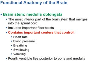
Medulla Oblongata
The medulla oblongata is the most inferior part of the brain stem, merging into the spinal cord. It contains important centers that regulate heart rate, blood pressure, breathing, swallowing, and vomiting.
Cardiovascular center: Controls heart rate and blood pressure
Respiratory center: Controls breathing
Other centers: Control swallowing and vomiting
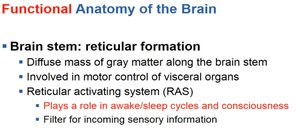
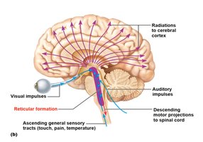
Reticular Formation
The reticular formation is a diffuse mass of gray matter along the brain stem, involved in motor control of visceral organs and regulating consciousness through the reticular activating system (RAS).
Motor control of visceral organs
RAS: Regulates sleep/wake cycles and consciousness
Filters incoming sensory information
Nervous Tissue: Neurons and Reflexes
Reflexes and Reflex Arcs
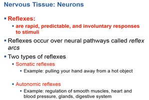
Reflexes are rapid, predictable, and involuntary responses to stimuli. They occur over neural pathways called reflex arcs, which are essential for protecting the body and maintaining homeostasis.
Somatic reflexes: Involve skeletal muscles (e.g., pulling hand away from hot object)
Autonomic reflexes: Involve smooth muscle, heart, glands (e.g., regulation of blood pressure)
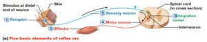
Elements of a Reflex Arc
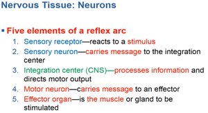
A reflex arc consists of five basic elements that work together to produce a rapid response to a stimulus.
Sensory receptor: Reacts to a stimulus
Sensory neuron: Carries message to the integration center
Integration center (CNS): Processes information and directs motor output
Motor neuron: Carries message to an effector
Effector organ: Muscle or gland that responds
Neuronal Communication: Synaptic Transmission
Structure of a Synapse
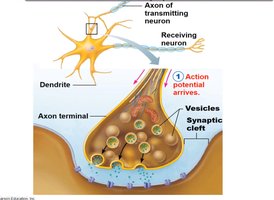
A synapse is the junction between two neurons where information is transmitted from one neuron to another. The axon terminal of the transmitting neuron communicates with the dendrite of the receiving neuron via the synaptic cleft.
Axon terminal: End of the transmitting neuron
Synaptic cleft: Small gap between neurons
Vesicles: Contain neurotransmitters
Steps of Synaptic Transmission
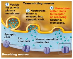
Neuronal communication across a synapse involves several key steps:
Action potential arrives at the axon terminal.
Vesicle fuses with the plasma membrane, releasing neurotransmitter into the synaptic cleft.
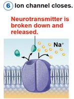
Neurotransmitter binds to receptors on the receiving neuron's membrane, opening ion channels.
Neurotransmitter is broken down and released, closing the ion channel and ending the signal.
Example: Acetylcholine is a neurotransmitter that binds to receptors on muscle cells, causing muscle contraction. After its action, acetylcholine is broken down by the enzyme acetylcholinesterase, terminating the signal.
Additional info: The process of synaptic transmission is essential for all neural communication, including reflexes, sensory perception, and voluntary movement.
