 Back
BackFunctional Anatomy of the Brain: Structure and Organization of the Central Nervous System
Study Guide - Smart Notes
 Tailored notes based on your materials, expanded with key definitions, examples, and context.
Central Nervous System (CNS)
Overview of Brain Regions
The central nervous system (CNS) consists of the brain and spinal cord. The brain is divided into several major regions, each with specialized functions essential for sensory processing, motor control, and higher cognitive activities.
Cerebral Hemispheres: The largest part of the brain, responsible for higher brain functions such as thought, voluntary movement, language, reasoning, and perception.
Diencephalon: Contains structures such as the thalamus, hypothalamus, and epithalamus, which are involved in sensory relay, autonomic control, and endocrine functions.
Brain Stem: Connects the brain to the spinal cord and controls vital life functions such as breathing, heart rate, and blood pressure.
Cerebellum: Coordinates voluntary movements, balance, and posture.
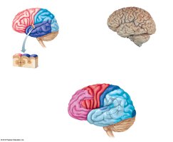
Cerebral Hemispheres
Surface Anatomy and Lobes
The cerebral hemispheres are paired structures that form the superior part of the brain. They are characterized by a highly folded surface, increasing the area for cortical neurons.
Gyri: Elevated ridges on the brain surface.
Sulci: Shallow grooves separating gyri.
Fissures: Deeper grooves that separate large regions of the brain.
Lobes: Named after the cranial bones that overlie them (frontal, parietal, temporal, occipital).
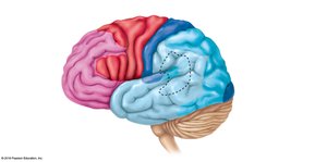
Cerebral Cortex: Functional Areas
The cerebral cortex is the outer layer of gray matter responsible for conscious thought, sensation, and voluntary movement. It is divided into several functional areas:
Primary Somatic Sensory Area: Located in the parietal lobe, posterior to the central sulcus; receives sensory input from the body (pain, temperature, touch).
Primary Motor Area: Located anterior to the central sulcus in the frontal lobe; controls voluntary movements of skeletal muscles.
Special Senses Areas: Visual area (occipital lobe), auditory area (temporal lobe), olfactory area (temporal lobe), gustatory area (taste).
Broca’s Area: Motor speech area, usually in the left hemisphere; involved in speech production.
Association Areas: Integrate and interpret information (anterior association area for judgment and problem-solving, posterior association area for language and spatial awareness).

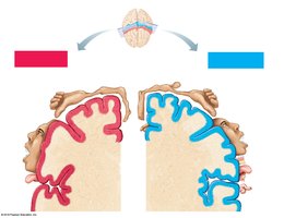
Homunculus Maps
The sensory homunculus and motor homunculus are spatial representations of the body within the primary somatic sensory and motor cortices, respectively. These maps illustrate the amount of cortex devoted to each body region, reflecting the density of sensory receptors or motor units.
The left side of the sensory cortex receives input from the right side of the body and vice versa.
Regions with fine motor control or high sensory acuity (e.g., hands, face) occupy larger cortical areas.
Cerebral White Matter and Basal Nuclei
Cerebral White Matter
White matter consists of myelinated fiber tracts beneath the gray matter cortex. These tracts are classified as:
Commissural fibers: Connect corresponding areas of the two hemispheres (e.g., corpus callosum).
Association fibers: Connect different parts of the same hemisphere.
Projection fibers: Connect the cerebrum with lower brain regions and the spinal cord.
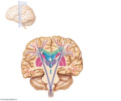
Basal Nuclei
The basal nuclei are clusters of gray matter deep within the cerebral white matter. They help regulate voluntary motor activities by modifying instructions sent from the primary motor cortex to skeletal muscles.
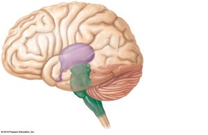
Diencephalon
Major Structures
The diencephalon is located above the brain stem and is surrounded by the cerebral hemispheres. It consists of three main structures:
Thalamus: Acts as a relay station for sensory impulses ascending to the cerebral cortex; directs impulses to the appropriate cortical area for processing.
Hypothalamus: Regulates autonomic functions such as body temperature, water balance, and metabolism; controls the pituitary gland and is involved in emotional responses (limbic system).
Epithalamus: Contains the pineal gland (an endocrine organ) and the choroid plexus (produces cerebrospinal fluid).
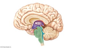
Brain Stem
Structure and Function
The brain stem connects the brain to the spinal cord and is responsible for many automatic functions necessary for survival. It is composed of three main parts:
Midbrain: Contains fiber tracts and reflex centers for vision and hearing (corpora quadrigemina).
Pons: Contains fiber tracts and nuclei involved in the control of breathing.
Medulla Oblongata: The most inferior part; contains centers that regulate heart rate, blood pressure, breathing, swallowing, and vomiting.

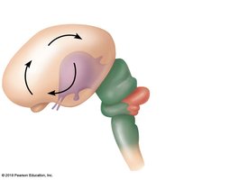
Cerebellum
Structure and Function
The cerebellum is located under the occipital lobes of the cerebrum and has two hemispheres with a highly folded surface. It coordinates voluntary movements, maintains balance and posture, and ensures smooth, precise timing of skeletal muscle activity.
Summary Table: Major Brain Regions and Functions
Region | Main Structures | Primary Functions |
|---|---|---|
Cerebral Hemispheres | Cortex, white matter, basal nuclei | Conscious thought, voluntary movement, sensory perception, language |
Diencephalon | Thalamus, hypothalamus, epithalamus | Sensory relay, autonomic regulation, endocrine control |
Brain Stem | Midbrain, pons, medulla oblongata | Vital centers for heart rate, breathing, reflexes |
Cerebellum | Two hemispheres, cortex, white matter | Coordination of movement, balance, posture |
