 Back
BackFunctional Divisions and Organization of the Peripheral Nervous System (PNS): Somatic and Autonomic Nervous Systems
Study Guide - Smart Notes
 Tailored notes based on your materials, expanded with key definitions, examples, and context.
Functional Divisions of the Peripheral Nervous System
Overview of the PNS
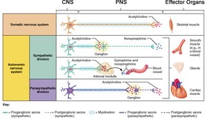
The Peripheral Nervous System (PNS) consists of all nervous tissue outside the central nervous system (CNS). It is functionally divided into the Somatic Nervous System (SNS) and the Autonomic Nervous System (ANS), each with distinct roles and effectors.
Somatic Nervous System (SNS): Controls voluntary movements of skeletal muscles.
Autonomic Nervous System (ANS): Regulates involuntary functions, including smooth muscle, cardiac muscle, and glandular activity.
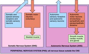
Somatic Nervous System (SNS)
Effectors and Control
The SNS is responsible for voluntary control of skeletal muscles. It involves a single motor neuron pathway from the CNS to the effector muscle.
Effectors: Skeletal muscles
Control: Voluntary (conscious) control
Motor Neurons: Large, fast, thickly myelinated Type A nerve fibers
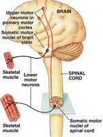
Organization of the Somatic Nervous System
The main control center for the SNS is the primary motor cortex of the cerebrum. The lower motor neuron extends from the CNS directly to the skeletal muscle effector.
Upper motor neurons: Located in the primary motor cortex or brainstem
Lower motor neurons: Extend from the CNS to the skeletal muscle
Autonomic Nervous System (ANS)
Effectors and Control
The ANS regulates involuntary activities of smooth muscle, cardiac muscle, and glands. It is divided into the sympathetic and parasympathetic divisions, which often have opposing effects on target organs.
Effectors: Smooth muscle, cardiac muscle, glands
Control: Involuntary (subconscious) control
Motor Neurons: Two-neuron chain (preganglionic and postganglionic neurons)
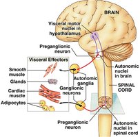
Organization of the Autonomic Nervous System
The ANS uses a two-neuron pathway: a preganglionic neuron (originating in the CNS) synapses with a postganglionic neuron in an autonomic ganglion outside the CNS. The postganglionic neuron then innervates the effector.
Control center: Hypothalamus
Preganglionic neuron: Originates in CNS, synapses in autonomic ganglia
Postganglionic neuron: Extends to effector
Somatic vs. Autonomic Nervous System: Key Differences
Effectors: SNS targets skeletal muscle; ANS targets smooth muscle, cardiac muscle, and glands.
Control: SNS is voluntary; ANS is involuntary.
Neural Pathway: SNS uses a single neuron; ANS uses a two-neuron chain.
Nerve Fiber Types: SNS uses Type A fibers; ANS uses Type B (preganglionic) and Type C (postganglionic) fibers.
Divisions of the Autonomic Nervous System
Sympathetic Division (Thoracolumbar Division)
The sympathetic division prepares the body for heightened activity ("fight or flight"). Preganglionic neurons originate in the thoracic and lumbar regions (T1–L2) of the spinal cord. Sympathetic ganglia are located near the spinal cord.
Sympathetic chain (paravertebral) ganglia: Innervate head, limbs, thoracic cavity
Prevertebral (collateral) ganglia: Innervate abdominopelvic organs
Preganglionic fibers: Short
Postganglionic fibers: Long

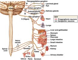
Parasympathetic Division (Craniosacral Division)
The parasympathetic division conserves energy and promotes maintenance functions ("rest and digest"). Preganglionic neurons originate in brain nuclei of cranial nerves III, VII, IX, X and sacral spinal cord segments. Parasympathetic ganglia are located near or within target organs.
Terminal (intramural) ganglia: Located near or inside effectors
Preganglionic fibers: Long
Postganglionic fibers: Short
Somatic & Autonomic Nervous System Organization: Comparative Overview
This table summarizes the organization, neurotransmitters, and effectors of the somatic and autonomic nervous systems.
System | Neural Pathway | Neurotransmitter(s) | Effector(s) |
|---|---|---|---|
Somatic | Single neuron (CNS to muscle) | Acetylcholine (ACh) | Skeletal muscle |
Autonomic - Sympathetic | Two neurons (preganglionic & postganglionic) | ACh (preganglionic), Norepinephrine (NE) or Epinephrine (E) (postganglionic) | Smooth muscle, cardiac muscle, glands |
Autonomic - Parasympathetic | Two neurons (preganglionic & postganglionic) | ACh (both neurons) | Smooth muscle, cardiac muscle, glands |
Neurotransmitters and Receptors in the PNS
Somatic Motor Neurons
Release acetylcholine (ACh) at neuromuscular junctions
Effect is always excitatory, causing muscle contraction
Autonomic Motor Neurons
All preganglionic neurons (both divisions) release ACh
Postganglionic parasympathetic neurons release ACh
Most postganglionic sympathetic neurons release norepinephrine (NE)
Response (excitation or inhibition) depends on receptor type
Cholinergic Neurons and Receptors
Cholinergic neurons: Release ACh
Nicotinic receptors: Found on skeletal muscle, all postganglionic neurons, adrenal medulla; always excitatory
Muscarinic receptors: Found on all parasympathetic targets and some sympathetic targets; effect can be excitatory or inhibitory
Adrenergic Neurons and Receptors
Adrenergic neurons: Release norepinephrine (NE)
Adrenergic receptors: Alpha (α) and beta (β) types
α1 and β1 receptors: Generally excitatory
α2 and β2 receptors: Generally inhibitory
Heart: β1 receptors increase heart rate and contraction
Respiratory airways: β2 receptors relax smooth muscle and dilate airways
Sympathetic vs. Parasympathetic Effects on Target Organs
The following table summarizes the effects of sympathetic and parasympathetic stimulation on various organs:
Target Organ | Parasympathetic Effects | Sympathetic Effects |
|---|---|---|
Blood vessels | No effect | Dilation to skeletal muscles & heart; constriction to viscera |
Salivary glands | Stimulates watery salivation | Inhibits watery salivation |
Sweat glands | No effect | Stimulates sweating |
Genitals | Stimulates erection | Causes glandular secretion, contraction, & ejaculation |
GI Tract | Increases motility & gland secretion; dilates sphincters | Decreases motility & gland secretion; constricts sphincters |
Heart | Decreases heart rate, force of contraction, & BP | Increases heart rate, force of contraction, & BP |
Respiratory Airways | Constricts bronchioles | Dilates bronchioles |
Eye (pupil) | Constricts pupil | Dilates pupil |
Dual Innervation and Autonomic Tone
Most visceral organs receive dual innervation from both sympathetic and parasympathetic divisions, allowing for precise regulation. Effects are typically antagonistic, but some structures (e.g., sweat glands, adrenal medulla, arrector pili muscles, most blood vessels) receive only sympathetic input.
Extent and Duration of Autonomic Activity
Sympathetic Activity
Extensive divergence: Each preganglionic neuron may synapse with 30+ postganglionic neurons, causing widespread effects.
Effects of NE last several seconds; hormonal release from adrenal medulla prolongs effects.
Parasympathetic Activity
Limited divergence: Each preganglionic neuron synapses with few postganglionic neurons, leading to localized effects.
Effects at muscarinic receptors are brief due to rapid inactivation of ACh by acetylcholinesterase (AChE).
