 Back
BackFundamentals of the Nervous System and Nervous Tissue: Structured Study Notes
Study Guide - Smart Notes
 Tailored notes based on your materials, expanded with key definitions, examples, and context.
Functions of the Nervous System
Overview of Nervous System Functions
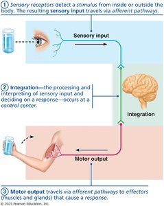
The nervous system is the master controlling and communicating system of the body. It operates through rapid and specific electrical and chemical signals, resulting in almost immediate responses. The nervous system performs three main functions:
Sensory Input: Gathering information from sensory receptors about internal and external changes.
Integration: Processing and interpretation of sensory input, deciding on a response.
Motor Output: Activation of effector organs (muscles and glands) to produce a response.
Organization of the Nervous System
Central and Peripheral Nervous Systems
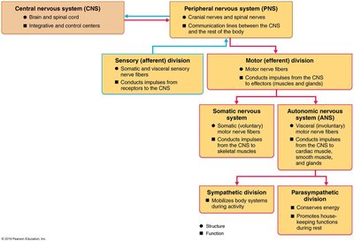
The nervous system is divided into two principal parts:
Central Nervous System (CNS): Composed of the brain and spinal cord, located in the dorsal body cavity. It serves as the integration and control center, interpreting sensory input and dictating motor output.
Peripheral Nervous System (PNS): Consists mainly of nerves extending from the brain and spinal cord. It connects the CNS to the rest of the body via spinal and cranial nerves.
Functional Divisions of the PNS
The PNS is further divided into:
Sensory (Afferent) Division:
Somatic sensory fibers: Convey impulses from skin, skeletal muscles, and joints to the CNS.
Visceral sensory fibers: Convey impulses from visceral organs to the CNS.
Motor (Efferent) Division:
Transmits impulses from the CNS to effector organs (muscles and glands).
Subdivided into the Somatic Nervous System (voluntary control of skeletal muscles) and the Autonomic Nervous System (involuntary control of smooth muscle, cardiac muscle, and glands).
The Autonomic Nervous System is further divided into Sympathetic and Parasympathetic subdivisions, which work in opposition to regulate body functions.
Nervous Tissue Cells
Principal Cell Types
Nervous tissue consists of two main cell types:
Neuroglia (Glial Cells): Small cells that surround and support neurons.
Neurons (Nerve Cells): Excitable cells that transmit electrical signals.
Neuroglia of the CNS
Four main types of neuroglia support CNS neurons:
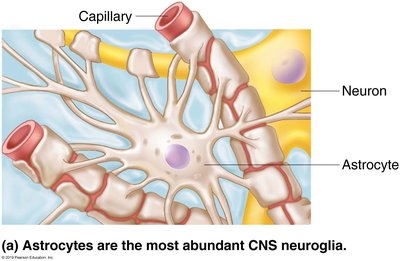
Astrocytes: Most abundant, versatile, and highly branched glial cells. They support and brace neurons, regulate exchanges between capillaries and neurons, control the chemical environment, respond to nerve impulses, and participate in information processing.
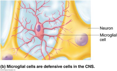
Microglial Cells: Small, ovoid cells with thorny processes. They monitor neurons, migrate toward injured neurons, and can transform to phagocytize microorganisms and neuronal debris.
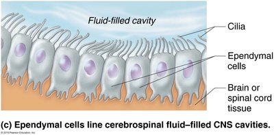
Ependymal Cells: May be ciliated; line the central cavities of the brain and spinal cord. Their cilia help circulate cerebrospinal fluid (CSF), and they form a permeable barrier between CSF and tissue fluid.
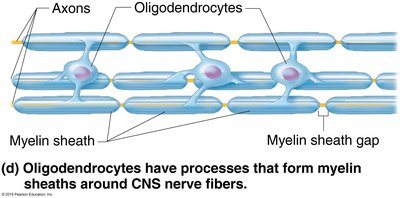
Oligodendrocytes: Branched cells whose processes wrap CNS nerve fibers, forming insulating myelin sheaths in thicker nerve fibers.
Neuroglia of the PNS
Two major neuroglia are found in the PNS:
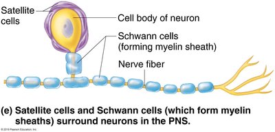
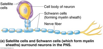
Satellite Cells: Surround neuron cell bodies in the PNS, functioning similarly to astrocytes.
Schwann Cells (Neurolemmocytes): Surround all peripheral nerve fibers and form myelin sheaths in thicker nerve fibers. They are vital for the regeneration of damaged peripheral nerve fibers.
Neurons: Structure and Function
Neuron Cell Body
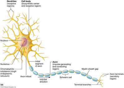
The neuron cell body is the biosynthetic center, synthesizing proteins, membranes, and chemicals. It contains a spherical nucleus with a nucleolus and may contain pigments. The plasma membrane is part of the receptive region, receiving input from other neurons.
Nuclei: Clusters of neuron cell bodies in the CNS.
Ganglia: Clusters of neuron cell bodies in the PNS.
Neuron Processes
Neurons have armlike processes extending from the cell body:
Dendrites: Receptive (input/afferent) regions that convey incoming messages as graded potentials.
Axon: Conducting region that generates and transmits nerve impulses. Axons may branch (axon collaterals) and end in axon terminals, which secrete neurotransmitters.
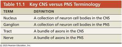
Key CNS vs. PNS Terminology
Term | Definition |
|---|---|
Nucleus | A collection of neuron cell bodies in the CNS |
Ganglion | A collection of neuron cell bodies in the PNS |
Tract | A bundle of axons in the CNS |
Nerve | A bundle of axons in the PNS |
Myelin Sheath
The myelin sheath is composed of myelin, a whitish, protein-lipid substance. Its functions include protecting and electrically insulating the axon and increasing the speed of nerve impulse transmission.
Myelinated fibers: Segmented sheath surrounds most long or large-diameter axons.
Myelination in the PNS: Schwann cells form myelin sheaths; gaps between cells are called nodes of Ranvier.
Myelination in the CNS: Oligodendrocytes form myelin sheaths; each cell can wrap up to 60 axons. White matter consists of myelinated fibers, while gray matter is mostly neuron cell bodies and nonmyelinated fibers.
Classification of Neurons
Structural Classification
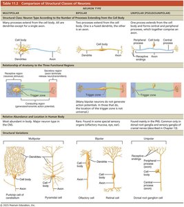
Neurons are classified by the number of processes:
Multipolar: Three or more processes (one axon, others dendrites); most common in CNS.
Bipolar: Two processes (one axon, one dendrite); rare, found in retina and olfactory mucosa.
Unipolar: One T-like process (two axons); also called pseudounipolar, associated with sensory receptors.
Functional Classification
Neurons are grouped by the direction in which nerve impulse travels relative to the CNS:
Sensory (Afferent): Transmit impulses from sensory receptors toward CNS; almost all are unipolar, cell bodies in PNS ganglia.
Motor (Efferent): Carry impulses from CNS to effectors; multipolar, most cell bodies in CNS.
Interneurons (Association Neurons): Lie between motor and sensory neurons, shuttle signals through CNS pathways; most are entirely within CNS.
Membrane Potentials and Signal Transmission
Resting Membrane Potential
Neurons have a resting membrane potential (RMP), which can rapidly change. Changes in membrane potential are used as signals to receive, integrate, and send information.
Depolarization: Decrease in membrane potential (moves toward zero and above); increases probability of producing an action potential.
Hyperpolarization: Increase in membrane potential (moves away from zero); decreases probability of producing an action potential.
Graded Potentials
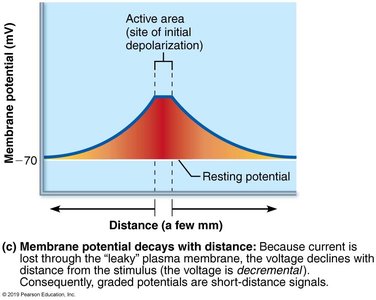
Graded potentials are short-lived, localized changes in membrane potential. They are triggered by stimuli that open gated ion channels and result in depolarization or hyperpolarization.
Receptor Potential: Graded potentials in receptors of sensory neurons.
Postsynaptic Potential: Graded potentials in neurons.
Graded potentials dissipate quickly and decay, serving as signals only over short distances.
Key Equations
Resting Membrane Potential Equation: Where:
= membrane potential
= gas constant
= temperature
= Faraday's constant
= extracellular potassium concentration
= intracellular potassium concentration
Summary Table: Structural Classes of Neurons
Neuron Type | Processes | Location |
|---|---|---|
Multipolar | Three or more (one axon, others dendrites) | Most common in CNS |
Bipolar | Two (one axon, one dendrite) | Retina, olfactory mucosa |
Unipolar | One T-like process (two axons) | Sensory receptors, PNS |
