 Back
BackFundamentals of the Nervous System and Nervous Tissue – Study Notes
Study Guide - Smart Notes
 Tailored notes based on your materials, expanded with key definitions, examples, and context.
Fundamentals of the Nervous System and Nervous Tissue
Functions of the Nervous System
The nervous system is responsible for controlling and integrating all body activities. It accomplishes this through three main functions:
Sensory Input: Collecting information from sensory receptors about internal and external changes.
Integration: Processing and interpreting sensory input to determine an appropriate response.
Motor Output: Activating effector organs (muscles and glands) to produce a response.
Organization of the Nervous System
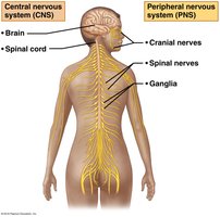
The nervous system is structurally and functionally divided into two main parts:
Central Nervous System (CNS): Consists of the brain and spinal cord; responsible for integration and command.
Peripheral Nervous System (PNS): Consists of cranial nerves, spinal nerves, and ganglia; connects the CNS to the rest of the body.
PNS Organization
The Peripheral Nervous System is further divided based on function:
Sensory (Afferent) Division: Transmits information from sensory receptors to the CNS.
Somatic sensory fibers: Carry information from skin, skeletal muscles, and joints.
Visceral sensory fibers: Carry information from visceral organs (e.g., stomach, intestines).
Motor (Efferent) Division: Transmits commands from the CNS to effector organs.
Somatic Nervous System: Controls voluntary movements of skeletal muscles.
Autonomic Nervous System: Controls involuntary actions of smooth muscle, cardiac muscle, and glands.
Sympathetic Division: Mobilizes body systems during activity ("fight or flight").
Parasympathetic Division: Conserves energy and promotes "rest and digest" functions.
Nervous Tissue Cells
Neuroglia of the CNS
Neuroglia (glial cells) are supporting cells in the CNS with specialized functions:
Astrocytes: Most abundant; regulate the chemical environment, guide neuron development, and support synapse formation.
Microglial Cells: Act as immune defense cells, protecting neurons from pathogens and injury.
Ependymal Cells: Line cavities of the brain and spinal cord; circulate cerebrospinal fluid (CSF) with cilia and form a permeable barrier.
Oligodendrocytes: Produce myelin sheaths around CNS nerve fibers, increasing conduction speed and providing insulation.
Neuroglia of the PNS
Satellite Cells: Surround neuron cell bodies in the PNS; function similarly to astrocytes.
Schwann Cells: Form myelin sheaths around peripheral nerve fibers and assist in nerve regeneration.
Neurons
General Characteristics
Neurons are the structural and functional units of the nervous system, specialized for rapid communication.
Longevity: Most neurons function for a lifetime.
Amitotic: Mature neurons do not divide.
High Metabolic Rate: Require continuous oxygen and glucose supply.
Neuron Structure
Cell Body (Soma): Contains the nucleus, rough ER (chromatophilic substance), neurofibrils, and pigment inclusions. In the CNS, clusters are called nuclei; in the PNS, ganglia.
Processes: Extensions from the cell body. In the CNS, bundles are called tracts; in the PNS, nerves.
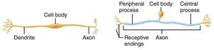
Dendrites: Receive signals and transmit them toward the cell body (graded potentials).
Axons: Generate and conduct nerve impulses away from the cell body; may have branches (collaterals) and end in axon terminals.
Myelination
PNS Myelination: Schwann cells wrap around axons, forming myelin sheaths with gaps called nodes of Ranvier. Myelination increases conduction speed.
CNS Myelination: Oligodendrocytes form myelin sheaths. White matter contains myelinated fibers; gray matter contains neuron cell bodies and unmyelinated fibers.
Conduction Velocity
Axon Diameter: Larger diameter axons conduct impulses faster.
Degree of Myelination: Myelinated axons conduct impulses via saltatory conduction (jumping from node to node), while unmyelinated axons use continuous conduction.
Classification of Neurons
Structural Classification
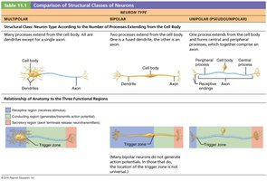
Neurons are classified by the number of processes extending from the cell body:
Type | Processes | Location/Function |
|---|---|---|
Multipolar | 3+ processes (1 axon, many dendrites) | Most common; major neuron type in CNS |
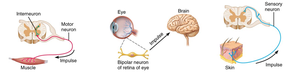
Bipolar | 2 processes (1 axon, 1 dendrite) | Rare; found in special sensory organs (e.g., retina) |
Unipolar (Pseudounipolar) | 1 process that splits into peripheral and central processes | Mainly sensory neurons in PNS |
Functional Classification
Sensory (Afferent) Neurons: Transmit impulses from sensory receptors toward the CNS; mostly unipolar, cell bodies in ganglia outside CNS.
Motor (Efferent) Neurons: Carry impulses from the CNS to effectors (muscles/glands); multipolar, cell bodies in CNS.
Interneurons (Association Neurons): Lie between sensory and motor neurons; most are within the CNS and are multipolar.
Synapses
Structure and Function of Synapses
A synapse is a junction that mediates information transfer from one neuron to another or from a neuron to an effector cell.
Presynaptic Neuron: Conducts impulses toward the synapse; sends information.
Postsynaptic Neuron: Receives the signal.
Types of Synapses:
Axodendritic: Between axon terminals of one neuron and dendrites of another.
Axosomatic: Between axon terminals and cell body.
Chemical Synapses
Specialized for release and reception of neurotransmitters.
Composed of an axon terminal (with synaptic vesicles) and a receptor region (on postsynaptic membrane), separated by a synaptic cleft.
Transmission involves conversion of electrical signal to chemical (neurotransmitter release), then back to electrical in the postsynaptic cell.
Electrical Synapses
Neurons are electrically coupled via gap junctions.
Allow direct flow of ions between cells, enabling rapid communication.
