 Back
BackFundamentals of the Nervous System and Nervous Tissue: Structured Study Notes
Study Guide - Smart Notes
 Tailored notes based on your materials, expanded with key definitions, examples, and context.
Fundamentals of the Nervous System and Nervous Tissue
Overview and Functions of the Nervous System
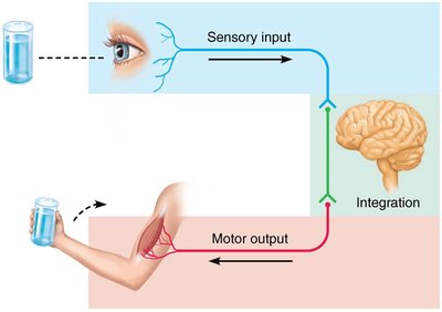
The nervous system is the master controlling and communicating system of the body. It utilizes electrical and chemical signals to coordinate rapid, specific responses. The nervous system's functions are divided into three overlapping stages:
Sensory Input: Information is gathered by sensory receptors about internal and external changes.
Integration: Processing and interpretation of sensory input occur in the central nervous system.
Motor Output: Activation of effector organs (muscles and glands) produces a response.
Example: When you see a glass of water (sensory input), your brain processes this information (integration), and your arm muscles move to pick up the glass (motor output).
Divisions of the Nervous System
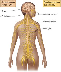
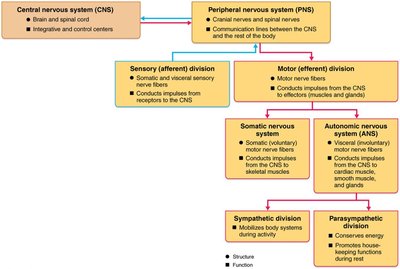
The nervous system is divided into two principal parts:
Central Nervous System (CNS): Composed of the brain and spinal cord, located in the dorsal body cavity. It serves as the integration and control center.
Peripheral Nervous System (PNS): Consists mainly of nerves that extend from the brain and spinal cord. It connects the CNS to the rest of the body via cranial and spinal nerves.
The PNS is further divided into:
Sensory (Afferent) Division: Transmits sensory information to the CNS.
Motor (Efferent) Division: Transmits commands from the CNS to effector organs.
Somatic Nervous System: Controls voluntary movements of skeletal muscles.
Autonomic Nervous System (ANS): Regulates involuntary functions (smooth muscle, cardiac muscle, glands) and is subdivided into sympathetic and parasympathetic divisions.
Cells of the Nervous System
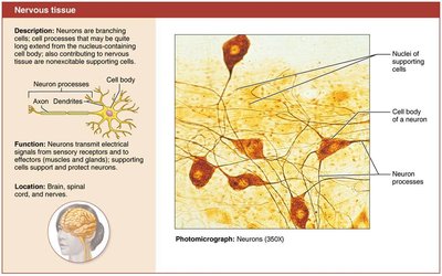
Nervous Tissue: Structure and Function
Nervous tissue consists of two principal cell types:
Neuroglia (Glial Cells): Support, protect, and insulate neurons.
Neurons (Nerve Cells): Excitable cells that transmit electrical signals.
Neuroglia of the CNS
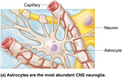
Astrocytes: Most abundant glial cells; support neurons, regulate exchanges between capillaries and neurons, guide neuron migration, and control the chemical environment.
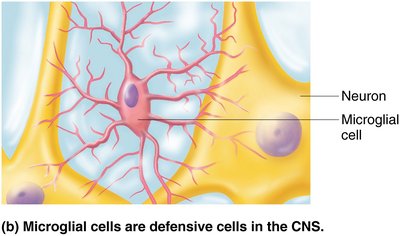
Microglia: Defensive cells; monitor neuron health and can transform to phagocytize microorganisms and debris.
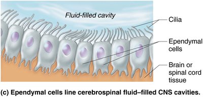
Ependymal Cells: Line cerebrospinal fluid-filled CNS cavities; may be ciliated to help circulate CSF.
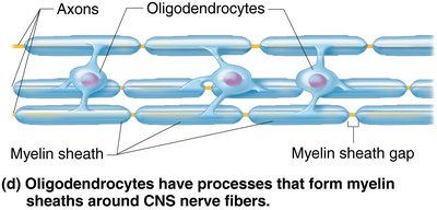
Oligodendrocytes: Form myelin sheaths around CNS nerve fibers, increasing impulse speed and insulation.
Neuroglia of the PNS
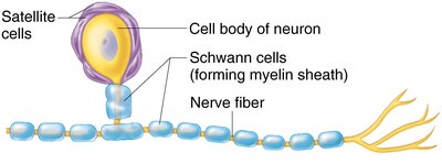
Satellite Cells: Surround neuron cell bodies in the PNS, similar in function to astrocytes.
Schwann Cells: Form myelin sheaths around peripheral nerve fibers and are vital for nerve regeneration.
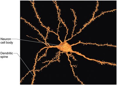
Neurons: Structure and Function
Neurons are the structural units of the nervous system. They are large, highly specialized cells that conduct impulses and possess the following characteristics:
Extreme Longevity: Can last a person's lifetime.
Amitotic: Most do not divide after maturity.
High Metabolic Rate: Require continuous supply of oxygen and glucose.
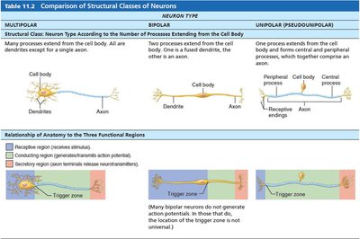
Structure: All neurons have a cell body (soma) and one or more processes (dendrites and axons).
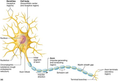
Neuron Processes: Dendrites and Axons
Dendrites: Receptive regions; convey incoming messages toward the cell body as graded potentials.
Axon: Conducting region; generates and transmits nerve impulses away from the cell body. Axons may be myelinated or nonmyelinated.
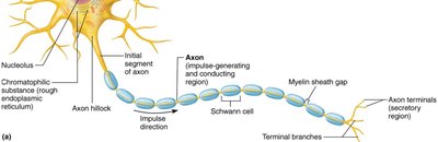
Myelination of Neurons
CNS Myelination: Oligodendrocytes form myelin sheaths; myelinated fibers transmit impulses faster.

PNS Myelination: Schwann cells wrap around axons in a jelly roll fashion, forming myelin sheaths. Myelin sheath gaps (nodes of Ranvier) are sites where axon collaterals can emerge.

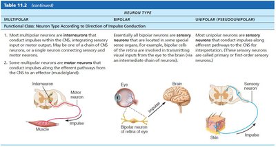
Structural and Functional Classification of Neurons
Multipolar: Three or more processes; most common in CNS.
Bipolar: Two processes; rare, found in retina and olfactory mucosa.
Unipolar: One T-like process; found mainly in PNS.
Neuron Type | Processes | Location |
|---|---|---|
Multipolar | Many dendrites, one axon | CNS |
Bipolar | One dendrite, one axon | Special sensory organs |
Unipolar | Single process | PNS |
Electrical Properties and Communication in Neurons
Resting Membrane Potential
Neurons have a resting membrane potential, typically around −70 mV. This is due to differences in ion concentrations and membrane permeability. The membrane is polarized, with the inside being more negative than the outside.
Generation and Changes in Membrane Potential
Depolarization: Membrane potential decreases (moves toward zero); increases probability of producing an impulse.
Hyperpolarization: Membrane potential increases (moves away from zero); decreases probability of producing an impulse.
Graded Potentials
Graded potentials are short-lived, localized changes in membrane potential, triggered by stimuli that open gated ion channels. The stronger the stimulus, the greater the voltage change and the farther the current flows.
Action Potentials
Action potentials are the principal means of long-distance neural communication. They involve a brief reversal of membrane potential (~100 mV) and occur only in muscle cells and axons of neurons. Action potentials do not decay over distance.
Resting State: All gated Na+ and K+ channels are closed.
Depolarization: Na+ channels open, Na+ rushes in, membrane potential rises.
Repolarization: Na+ channels inactivate, K+ channels open, K+ exits, membrane returns to resting potential.
Hyperpolarization: Some K+ channels remain open, causing a slight dip below resting voltage.
Threshold and All-or-None Phenomenon
For an axon to fire, depolarization must reach a threshold voltage. If threshold is reached, an action potential occurs completely; if not, it does not occur at all.
Propagation of Action Potentials
Action potentials are propagated along the axon, allowing transmission from origin to terminals. In myelinated axons, propagation is faster due to saltatory conduction.
Refractory Periods
Absolute Refractory Period: Neuron cannot trigger another AP; ensures one-way transmission.
Relative Refractory Period: Follows absolute period; only exceptionally strong stimulus can trigger AP.
Conduction Velocity
Axon Diameter: Larger-diameter fibers conduct impulses faster.
Degree of Myelination: Myelinated fibers conduct impulses much faster (saltatory conduction) than nonmyelinated fibers (continuous conduction).
The Synapse and Neural Circuitry
Synapses: Structure and Function
Synapses are junctions that mediate information transfer from one neuron to another or to an effector cell. There are two main types:
Chemical Synapse: Most common; involves release and reception of neurotransmitters.
Electrical Synapse: Less common; neurons are electrically coupled via gap junctions.
Information Transfer Across Chemical Synapses
Action potential arrives at axon terminal of presynaptic neuron.
Voltage-gated Ca2+ channels open, Ca2+ enters axon terminal.
Ca2+ entry causes synaptic vesicles to release neurotransmitter (exocytosis).
Neurotransmitter diffuses across synaptic cleft and binds to receptors on postsynaptic membrane.
Binding opens ion channels, creating graded potentials.
Neurotransmitter effects are terminated by reuptake, degradation, or diffusion.
Postsynaptic Potentials
Excitatory Postsynaptic Potentials (EPSPs): Depolarize the postsynaptic membrane, increasing likelihood of AP.
Inhibitory Postsynaptic Potentials (IPSPs): Hyperpolarize the postsynaptic membrane, decreasing likelihood of AP.
Summation of Synaptic Events
Temporal Summation: Rapid-fire impulses from one presynaptic neuron add together.
Spatial Summation: Simultaneous stimulation by multiple presynaptic neurons adds together.
Neurotransmitters: The Language of the Nervous System
Chemical Classes of Neurotransmitters
Acetylcholine (ACh): Memory and muscle function; degraded by acetylcholinesterase.
Biogenic Amines: Includes catecholamines (dopamine, epinephrine, norepinephrine) and indolamines (serotonin, histamine).
Amino Acids: Glutamate (excitatory), GABA and glycine (inhibitory).
Peptides: Substance P, endorphins (pain regulation).
Purines: Adenosine (caffeine blocks adenosine receptors).
Gases: Nitric oxide (NO), carbon monoxide (CO).
Endocannabinoids: THC (memory, learning, appetite).
Functional Classification of Neurotransmitters
Effects: Excitatory (depolarizing) or inhibitory (hyperpolarizing), depending on receptor type.
Actions: Direct (binds and opens ion channels) or indirect (acts through second messengers).
Types of Neurotransmitter Receptors
Channel-linked (Ligand-gated) Receptors: Immediate, brief effects; e.g., nicotinic ACh receptors.
G Protein–linked Receptors: Indirect, complex, slow, and prolonged effects; e.g., muscarinic ACh receptors.
Neural Integration and Circuitry
Neuronal Pools and Processing
Neuronal Pool: Functional groups of neurons that integrate incoming information and forward processed information.
Serial Processing: Input travels along one pathway; produces specific, anticipated responses (e.g., spinal reflex).
Parallel Processing: Input travels along several pathways; promotes higher-level mental functioning.
Types of Circuits in Neuronal Pools
Diverging Circuit: One input, many outputs; amplifies signal.
Converging Circuit: Many inputs, one output; concentrates signal.
Reverberating Circuit: Chain of neurons with feedback; controls rhythmic activities.
Parallel After-Discharge Circuit: Several pathways; produces bursts of impulses.
Additional info: These notes expand on the original lecture slides by providing definitions, examples, and structured tables for clarity. All included images directly reinforce the adjacent explanations and are strictly relevant to the topic.
