 Back
BackFundamentals of the Nervous System and Nervous Tissue: Study Notes
Study Guide - Smart Notes
 Tailored notes based on your materials, expanded with key definitions, examples, and context.
Fundamentals of the Nervous System and Nervous Tissue
Overview of the Nervous System
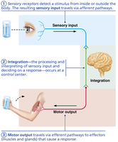
The nervous system is the primary control and communication system of the body. It uses electrical and chemical signals to coordinate rapid, specific responses to internal and external stimuli.
Sensory input: Information is gathered by sensory receptors about changes inside and outside the body.
Integration: The nervous system processes and interprets sensory input and decides what should be done at each moment.
Motor output: The nervous system activates effector organs (muscles and glands) to cause a response.
Divisions of the Nervous System
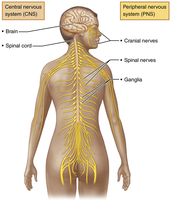
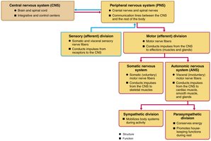
The nervous system is divided into two main parts: the central nervous system (CNS) and the peripheral nervous system (PNS).
Central Nervous System (CNS): Consists of the brain and spinal cord. It is the integration and control center.
Peripheral Nervous System (PNS): Consists of nerves that extend from the brain and spinal cord (cranial and spinal nerves). It serves as communication lines between the CNS and the rest of the body.
Organization of the Nervous System
The PNS is further divided into sensory (afferent) and motor (efferent) divisions. The motor division is subdivided into the somatic nervous system and the autonomic nervous system (ANS), which itself has sympathetic and parasympathetic divisions.
Cells of the Nervous System
Neurons
Neurons are the functional units of the nervous system. They are highly specialized cells that conduct electrical impulses. Key characteristics include:
Extreme longevity (can live a lifetime)
Amitotic (do not divide after maturity, with few exceptions)
High metabolic rate (require continuous oxygen and glucose)

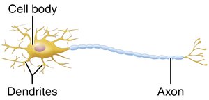
Structure of a Motor Neuron
Neurons have several processes extending from the cell body:
Dendrites: Receive incoming signals and convey them toward the cell body.
Axon: Conducts impulses away from the cell body to other neurons or effectors. The axon hillock is the cone-shaped area where the axon originates.
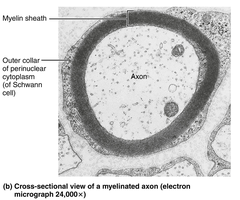
Myelin Sheath
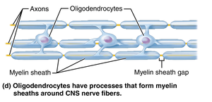
The myelin sheath is a white, fatty covering that insulates axons and increases the speed of nerve impulse transmission. Myelinated axons conduct impulses much faster than nonmyelinated axons.
Neuroglia (Glial Cells)
Neuroglia are supporting cells that protect, insulate, and support neurons. There are different types in the CNS and PNS:
Neuroglia in the CNS
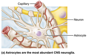
Astrocytes: Most abundant; support neurons, anchor them to capillaries, and help maintain the blood-brain barrier.
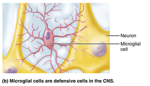
Microglial cells: Act as the main defense in the CNS; can transform into phagocytes to remove debris and pathogens.
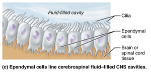
Ependymal cells: Line cerebrospinal fluid-filled cavities and help circulate CSF with their cilia.
Oligodendrocytes: Form myelin sheaths around CNS nerve fibers.
Neuroglia in the PNS
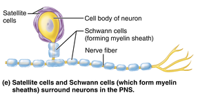
Satellite cells: Surround neuron cell bodies in the PNS; function similarly to astrocytes.
Schwann cells: Surround all peripheral nerve fibers and form myelin sheaths in the PNS; similar to oligodendrocytes in function.
Classification of Neurons
Structural Classification
Multipolar neurons: Many processes (1 axon, many dendrites); most common in CNS.
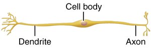
Bipolar neurons: Two processes (1 axon, 1 dendrite); rare, found in special senses.
Unipolar neurons: Single process that divides into two branches; mainly in PNS as sensory neurons.
Functional Classification
Sensory (afferent) neurons: Transmit impulses from sensory receptors toward the CNS.
Motor (efferent) neurons: Carry impulses from the CNS to effectors (muscles/glands).
Interneurons: Shuttle signals within the CNS; most abundant type.
Membrane Potentials and Nerve Impulses
Resting Membrane Potential
Neurons have a resting membrane potential of about -70 mV, generated by differences in ionic composition and membrane permeability. The sodium-potassium pump and selective ion channels maintain this potential.
Types of Signals
Graded potentials: Short-lived, localized changes in membrane potential; act as signals over short distances.
Action potentials: Long-distance signals of axons; do not decay with distance and follow the all-or-none principle.
Depolarization and Hyperpolarization
Depolarization: Membrane potential becomes less negative; increases likelihood of impulse generation.
Hyperpolarization: Membrane potential becomes more negative; decreases likelihood of impulse generation.
Synapses and Neurotransmitters
Synapses
A synapse is a junction where a neuron communicates with another cell. The presynaptic neuron sends the signal, and the postsynaptic neuron receives it. In the PNS, the postsynaptic cell may be a neuron, muscle, or gland cell.
Chemical Synapses
At chemical synapses, neurotransmitters are released from the presynaptic neuron and bind to receptors on the postsynaptic cell, causing graded potentials.
Postsynaptic Potentials
Excitatory postsynaptic potentials (EPSPs): Depolarize the postsynaptic membrane, increasing the chance of an action potential.
Inhibitory postsynaptic potentials (IPSPs): Hyperpolarize the postsynaptic membrane, decreasing the chance of an action potential.
Neurotransmitters
Neurotransmitters are chemical messengers that transmit signals across synapses. Over 50 have been identified, and they can be classified by structure and function:
Acetylcholine (ACh): Released at neuromuscular junctions; can be excitatory or inhibitory depending on receptor type.
Biogenic amines: Include catecholamines (dopamine, norepinephrine, epinephrine) and indolamines (serotonin, histamine).
Amino acids: Glutamate (excitatory), GABA and glycine (inhibitory).
Peptides: Substance P (pain mediator), endorphins (natural opiates).
Purines: ATP and adenosine (inhibitory in the brain).
Gases and lipids: Nitric oxide, carbon monoxide, hydrogen sulfide (act via G protein–coupled receptors).
Neurotransmitter Receptors
Channel-linked receptors: Ligand-gated ion channels; immediate and brief action.
G protein–linked receptors: Indirect, complex, slow, and often prolonged responses; cause widespread metabolic changes.
