 Back
BackChapter 11 - Fundamentals of the Nervous System & Nervous Tissue
Study Guide - Smart Notes
 Tailored notes based on your materials, expanded with key definitions, examples, and context.
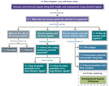
Introduction to the Nervous System
Overview and Organization
The nervous system is the master control and communication system of the body. It uses electrical signals along neurons and chemical signals at synapses to coordinate body functions. The nervous system is organized into the central nervous system (CNS) and peripheral nervous system (PNS), each with specialized roles in processing and transmitting information.
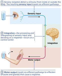
Functions of the Nervous System
Sensory Input: Sensory receptors detect stimuli from inside or outside the body and send information to the CNS via afferent pathways.
Integration: The CNS processes and interprets sensory input, deciding on an appropriate response.
Motor Output: The CNS sends signals via efferent pathways to effectors (muscles or glands) to produce a response.
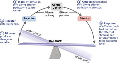
Homeostatic Control and Feedback
The nervous system maintains homeostasis through feedback mechanisms involving receptors, control centers, and effectors. Negative feedback loops are common, reducing the effect of a stimulus to restore balance.
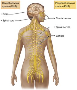
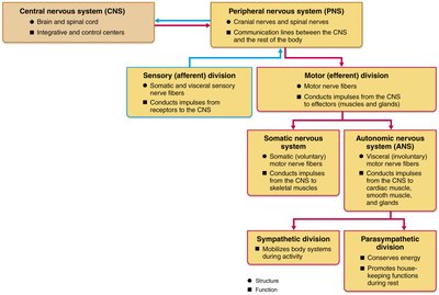
Anatomical Divisions of the Nervous System
Central and Peripheral Nervous Systems
Central Nervous System (CNS): Consists of the brain and spinal cord; responsible for integration and control.
Peripheral Nervous System (PNS): Includes cranial nerves, spinal nerves, and ganglia; connects the CNS to the rest of the body.
Functional Divisions of the PNS
Sensory (Afferent) Division: Transmits sensory information to the CNS.
Motor (Efferent) Division: Transmits commands from the CNS to effectors.
Somatic Nervous System: Controls voluntary movements (skeletal muscle).
Autonomic Nervous System (ANS): Controls involuntary functions (smooth muscle, cardiac muscle, glands).
Sympathetic Division: Mobilizes body systems during activity ("fight or flight").
Parasympathetic Division: Conserves energy and promotes housekeeping functions during rest ("rest and digest").
Cells of the Nervous System
Neuroglia (Glial Cells)
Neuroglia are supporting cells that maintain the environment around neurons, provide insulation, and assist in repair. They are classified by location and function:
CNS Neuroglia:
Astrocytes: Star-shaped, most abundant; support neurons and regulate nutrient exchange.
Microglial Cells: Immune cells that act as macrophages in the CNS.
Ependymal Cells: Line brain ventricles and spinal cord; circulate cerebrospinal fluid (CSF) with cilia.
Oligodendrocytes: Form myelin sheaths around CNS axons for insulation.
PNS Neuroglia:
Satellite Cells: Surround neuron cell bodies in the PNS; similar function to astrocytes.
Schwann Cells: Form myelin sheaths in the PNS and assist in nerve regeneration.
Neurons: Structure and Classification
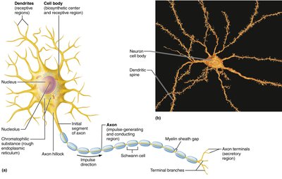
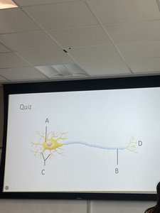
Neuron Structure
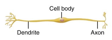
Neurons are excitable cells that transmit electrical signals. Key parts include:
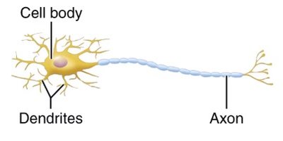
Cell Body (Soma): Contains the nucleus and organelles; biosynthetic center.
Dendrites: Receive incoming signals and convey them toward the cell body.
Axon: Conducts impulses away from the cell body to other neurons or effectors.
Myelin Sheath: Insulating layer that increases the speed of impulse transmission.
Nodes of Ranvier: Gaps in the myelin sheath where action potentials are regenerated.
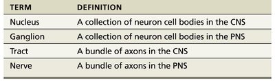
Terminology Table
TERM | DEFINITION |
|---|---|
Nucleus | A collection of neuron cell bodies in the CNS |
Ganglion | A collection of neuron cell bodies in the PNS |
Tract | A bundle of axons in the CNS |
Nerve | A bundle of axons in the PNS |
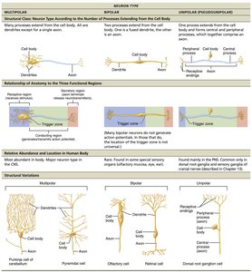
Structural Classification of Neurons
Multipolar Neurons: Many dendrites, one axon; most common in CNS.
Bipolar Neurons: One dendrite, one axon; found in retina and olfactory epithelium.
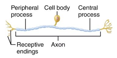
Unipolar (Pseudounipolar) Neurons: Single process that splits into peripheral and central branches; sensory neurons in PNS.
Functional Classification of Neurons
Sensory (Afferent) Neurons: Transmit impulses from sensory receptors to the CNS.
Motor (Efferent) Neurons: Carry impulses from the CNS to effectors (muscles/glands).
Interneurons: Connect sensory and motor neurons within the CNS; most abundant type.
Neurophysiology: Membrane Potentials and Signals
Ohm’s Law and Electrical Principles
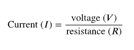
Neurons use electrical currents to transmit information. Ohm’s Law describes the relationship between current, voltage, and resistance:
Current (I): Flow of electrical charge (ions).
Voltage (V): Potential energy difference between two points.
Resistance (R): Hindrance to charge flow.
Ohm’s Law equation:
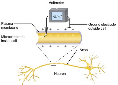
Resting Membrane Potential
The resting membrane potential is typically -70 mV in neurons, maintained by ion gradients and selective permeability of the plasma membrane. Key contributors include:
K+ Leak Channels: Allow K+ to diffuse out, making the inside more negative.
Na+ Leak Channels: Allow some Na+ to enter.
Na+/K+ Pump: Actively transports 3 Na+ out and 2 K+ in, maintaining gradients.
Electrochemical Gradients
Concentration Gradient: Ions move from high to low concentration.
Electrical Gradient: Opposite charges attract, influencing ion movement.
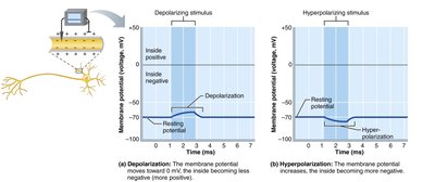
Changes in Membrane Potential
Depolarization: Membrane potential becomes less negative (more positive).
Hyperpolarization: Membrane potential becomes more negative.
Graded Potentials and Action Potentials
Graded Potentials
Graded potentials are short-lived, localized changes in membrane potential. They can be excitatory (EPSPs) or inhibitory (IPSPs) and are important for initiating action potentials if they reach threshold at the axon hillock.
EPSP: Depolarization that brings the membrane potential closer to threshold.
IPSP: Hyperpolarization that moves the membrane potential further from threshold.
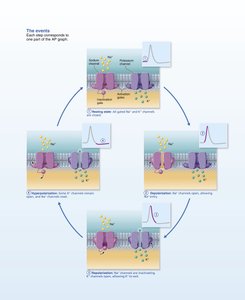
Action Potentials
Action potentials are long-distance signals of axons. They are all-or-none events that occur if the membrane potential reaches threshold (about -55 mV). The sequence includes depolarization (Na+ influx), repolarization (K+ efflux), and hyperpolarization.
Threshold: The critical level to which a membrane potential must be depolarized to initiate an action potential.
Refractory Periods: Absolute (no new AP possible) and relative (stronger stimulus needed for AP).
Comparison Table: Graded vs. Action Potentials
Graded Potential (GP) | Action Potential (AP) | |
|---|---|---|
Function | Short-distance signaling; determines if AP is generated | Long-distance signaling; nerve impulse |
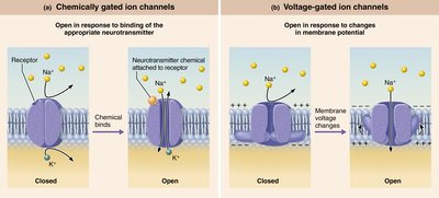
Initial Effect | Opens chemically gated channels | Opens voltage-gated channels |
Peak Membrane Potential | Depolarizes toward 0 mV or hyperpolarizes toward -90 mV | -70 mV to +30 mV |
Synapses and Neurotransmitters
Types of Synapses
Electrical Synapses: Use gap junctions for direct ion flow; synchronize activity (e.g., cardiac muscle).
Chemical Synapses: Most common; use neurotransmitters to transmit signals across a synaptic cleft; unidirectional.

Neurotransmitters
Excitatory: Glutamate, norepinephrine (NE)
Inhibitory: GABA
Modulatory: Serotonin (5-HT), dopamine, NE
Neurotransmitter Table (Selected Examples)
Neurotransmitter | Functional Class | Sites Where Secreted | Comments |
|---|---|---|---|
Acetylcholine (ACh) | Excitatory or inhibitory | CNS, PNS, neuromuscular junctions | Major neurotransmitter of the PNS; involved in muscle contraction |
Dopamine | Excitatory or inhibitory | CNS, some PNS | "Feel good" neurotransmitter; involved in reward and movement |
GABA | Generally inhibitory | CNS | Main inhibitory neurotransmitter in the brain |
Glutamate | Generally excitatory | CNS | Main excitatory neurotransmitter in the brain |
Summary
The nervous system is a complex network that integrates sensory input, processes information, and coordinates motor output. Neurons and neuroglia work together to maintain homeostasis and enable rapid communication throughout the body. Understanding the structure, function, and signaling mechanisms of nervous tissue is fundamental to the study of human physiology.
