 Back
Fundamentals of the Nervous System & Nervous Tissue: Structured Study Notes
Study Guide - Smart Notes
 Tailored notes based on your materials, expanded with key definitions, examples, and context.
Fundamentals of the Nervous System & Nervous Tissue
Nervous System Functions
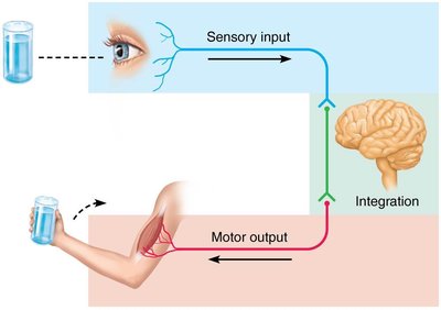
The nervous system is the master controlling and communicating system of the body. It operates through rapid and specific electrical and chemical signals, resulting in almost immediate responses. The nervous system has three overlapping functions:
Sensory Input: Information is gathered by sensory receptors about internal and external changes.
Integration: Processing and interpretation of sensory input occurs in the brain.
Motor Output: Activation of effector organs (muscles and glands) produces a response.
Example: When you see a glass of water, your sensory receptors send input to your brain, which integrates the information and sends motor output to your arm muscles to pick up the glass.
Nervous System Divisions
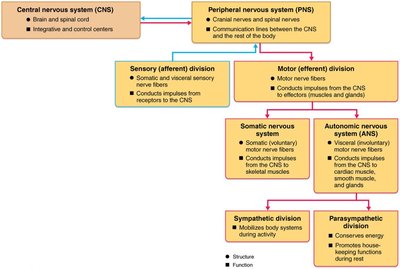
The nervous system is divided into two principal parts:
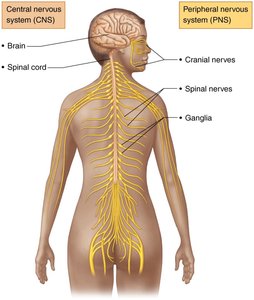
Central Nervous System (CNS): Consists of the brain and spinal cord, located in the dorsal body cavity. It serves as the integration and control center, interpreting sensory input and dictating motor output.
Peripheral Nervous System (PNS): The portion outside the CNS, mainly composed of nerves that extend from the brain and spinal cord. It includes spinal nerves (to and from the spinal cord) and cranial nerves (to and from the brain).
Organization of the Nervous System
The PNS is further divided into:
Sensory (Afferent) Division: Conducts impulses from sensory receptors to the CNS.
Motor (Efferent) Division: Conducts impulses from the CNS to effectors (muscles and glands).
The motor division includes:
Somatic Nervous System: Voluntary control of skeletal muscles.
Autonomic Nervous System (ANS): Involuntary control of smooth muscle, cardiac muscle, and glands. The ANS is subdivided into the sympathetic (mobilizes body systems) and parasympathetic (conserves energy) divisions.
Nervous Tissue: Cell Types
Nervous tissue consists of two principal cell types:
Neuroglia (Glial Cells): Small cells that surround and wrap delicate neurons.
Neurons (Nerve Cells): Excitable cells that transmit electrical signals.
Neuroglia of the CNS
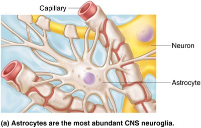
Astrocytes: Most abundant, versatile, and highly branched glial cells. They support and brace neurons, guide migration, control the chemical environment, and participate in information processing.
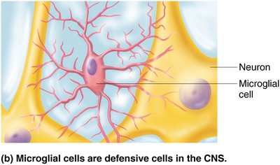
Microglial Cells: Small, ovoid cells with thorny processes. They migrate toward injured neurons and can transform to phagocytize microorganisms and neuronal debris.
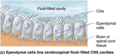
Ependymal Cells: Range in shape from squamous to columnar, may be ciliated. They line the central cavities of the brain and spinal column, forming a permeable barrier between cerebrospinal fluid (CSF) and tissue fluid.
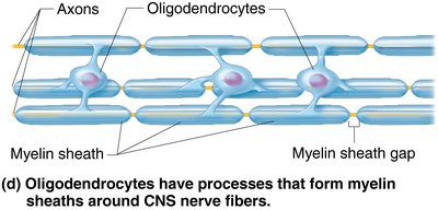
Oligodendrocytes: Branched cells whose processes wrap CNS nerve fibers, forming insulating myelin sheaths in thicker nerve fibers.
Neuroglia of the PNS
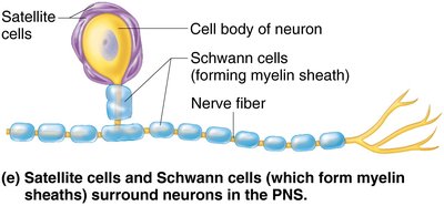
Satellite Cells: Surround neuron cell bodies in the PNS, functioning similarly to astrocytes in the CNS.
Schwann Cells: Surround all peripheral nerve fibers and form myelin sheaths in thicker nerve fibers. Vital to regeneration of damaged peripheral nerve fibers.
Neurons: Structure and Function
Neurons are the structural units of the nervous system. They are large, highly specialized cells that conduct impulses. Key characteristics include:
Extreme Longevity: Last a person's lifetime.
Amitotic: With few exceptions, they do not divide.
High Metabolic Rate: Require continuous supply of oxygen and glucose.
Structure: All have a cell body (soma) and one or more processes (dendrites and axons).
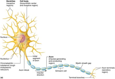
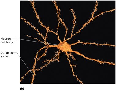
Neuron Cell Body (Soma)
Biosynthetic Center: Contains the nucleus and nucleolus.
Location: Most neuron cell bodies are in the CNS (called nuclei); in the PNS, they are called ganglia.
Neuron Processes
Dendrites: Receptive (input) regions of the neuron.
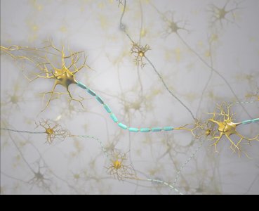
Axon: Conducting (output) region, generates and transmits nerve impulses to axon terminals, which secrete neurotransmitters.
The Myelin Sheath
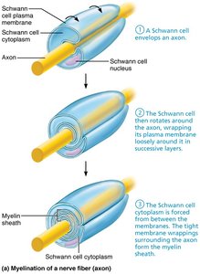
The myelin sheath is composed of myelin, a whitish, protein-lipid substance. Its functions include:
Protecting and electrically insulating the axon
Increasing the speed of nerve impulse transmission
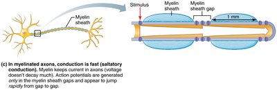
Myelinated fibers have segmented sheaths; non-myelinated fibers conduct impulses more slowly. In the PNS, myelin is formed by Schwann cells; in the CNS, by oligodendrocytes.
Classification of Neurons
Structural Classification
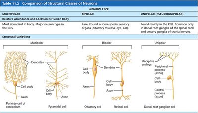
Neurons are classified based on the number and arrangement of their processes:
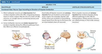
Multipolar: Many processes; most abundant in the body.
Bipolar: Two processes; found in special sensory organs.
Unipolar (Pseudounipolar): Single process; found mainly in the PNS.
Neuron Type | Location | Structural Variations |
|---|---|---|
Multipolar | Major neuron type in CNS | Pyramidal cell, Purkinje cell |
Bipolar | Special sensory organs | Olfactory cell, Retinal cell |
Unipolar | PNS | Dorsal root ganglion cell |
Functional Classification
Sensory (Afferent) Neurons: Transmit impulses from sensory receptors toward the CNS; almost all are unipolar.
Motor (Efferent) Neurons: Carry impulses from the CNS to effectors; multipolar.
Interneurons: Lie between motor and sensory neurons; shuttle signals through CNS pathways; most are entirely within the CNS.
Membrane Potentials and Basic Principles of Electricity
Neurons have a resting membrane potential and can rapidly change it. Key principles:
Opposite charges are attracted to each other.
Energy is required to keep opposite charges separated across a membrane.
Energy is liberated when charges move toward one another.
When separated, the system has potential energy.
Definitions:
Voltage: Measure of potential energy generated by separated charge (in volts or millivolts).
Current: Flow of electrical charge (ions) between two points.
Resistance: Hindrance to charge flow.
Ohm’s Law:
Role of Membrane Ion Channels
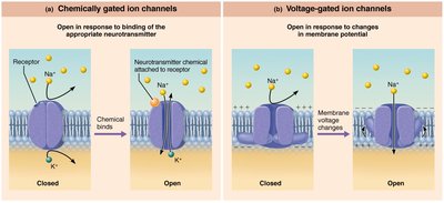
Large proteins serve as selective membrane ion channels. Types include:
Leakage (Nongated) Channels: Always open.
Gated Channels: Open/close in response to specific signals. Types: chemically gated, voltage-gated, mechanically gated.
Resting Membrane Potential (RMP)
The RMP of a resting neuron is approximately −70 mV. The cytoplasmic side is negatively charged relative to the outside, and the membrane is polarized. RMP is generated by:
Differences in ionic composition of intracellular and extracellular fluids
Differences in plasma membrane permeability
ECF has higher Na+; ICF has higher K+. K+ plays the most important role in membrane potential.
Changes in Membrane Potential
Membrane potential changes when ion concentrations or membrane permeability change. These changes produce:
Graded Potentials: Incoming signals operating over short distances.
Action Potentials: Long-distance signals of axons.
Terms:
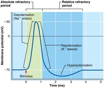
Depolarization: Decrease in membrane potential (moves toward zero and above).
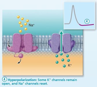
Hyperpolarization: Increase in membrane potential (away from zero).
Graded Potentials
Graded potentials are short-lived, localized changes in membrane potential. The stronger the stimulus, the more voltage changes and the farther current flows. They are triggered by stimuli that open gated ion channels and result in depolarization or hyperpolarization. Graded potentials dissipate quickly and decay.
Action Potentials (AP)
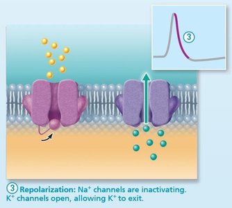
Action potentials are the principal way neurons send signals for long-distance communication. They occur only in muscle cells and axons of neurons, involving a brief reversal of membrane potential (~100 mV). APs do not decay over distance.
Threshold & All-or-None Phenomenon
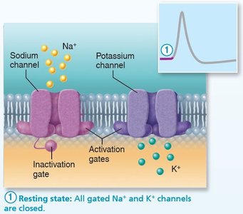
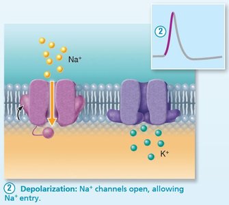
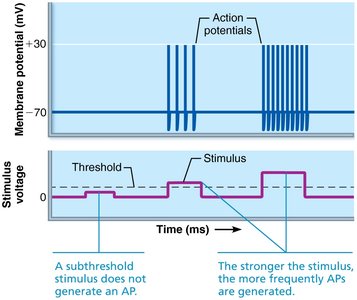
For an axon to "fire," depolarization must reach threshold voltage. At threshold, Na+ permeability increases, and the positive feedback cycle begins. An AP either happens completely or not at all. Propagation allows AP to be transmitted from origin down the entire axon length.
Coding for Stimulus Intensity
All action potentials are alike and independent of stimulus intensity. The CNS distinguishes between weak and strong stimuli by the frequency of impulses (number of APs per second). Higher frequencies indicate stronger stimuli.
Refractory Periods
The refractory period is the time during which a neuron cannot trigger another AP. Types:
Absolute Refractory Period: Time from opening of Na+ channels until resetting; ensures all-or-none event and one-way transmission.
Relative Refractory Period: Follows absolute period; most Na+ channels have returned to resting state, some K+ channels still open. Only exceptionally strong stimulus can trigger an AP.
Conduction Velocity
The rate of AP propagation depends on axon diameter and degree of myelination:
Continuous Conduction: Slow, occurs in nonmyelinated axons.
Saltatory Conduction: Fast, occurs only in myelinated axons; APs jump from gap to gap.
Nerve Fiber Classification
Group | Diameter | Myelination | Speed | Function |
|---|---|---|---|---|
Group A | Largest | Myelinated | 150 m/s | Somatic sensory and motor fibers |
Group B | Intermediate | Lightly myelinated | 15 m/s | ANS visceral motor and sensory fibers |
Group C | Smallest | Unmyelinated | 1 m/s | ANS visceral motor and sensory fibers |
Clinical Homeostatic Imbalance
Impaired AP impulse propagation can be caused by chemical and physical factors:
Local Anesthetics: Block voltage-gated Na+ channels.
Cold/Pressure: Interrupt blood circulation and oxygen delivery to neurons.
Multiple Sclerosis (MS): Autoimmune disease destroying CNS myelin sheaths, leading to hardened lesions (scleroses), slowed impulse conduction, and symptoms such as visual disturbances, weakness, and incontinence. Treatment involves drugs modifying immune system activity; high vitamin D may reduce risk.
References: Marieb, E. N., & Hoehn, K. (2023). Human anatomy and physiology (12th ed.). Pearson.
