 Back
Fundamentals of the Nervous System & Nervous Tissue: Synapses, Neurotransmitters, and Neural Integration
Study Guide - Smart Notes
 Tailored notes based on your materials, expanded with key definitions, examples, and context.
Structure and Function of Synapses
Introduction to Synapses
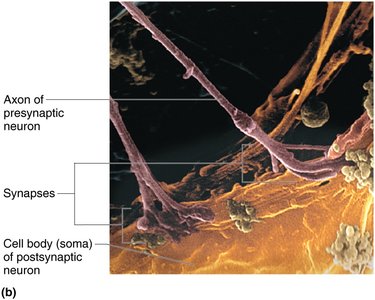
Synapses are specialized junctions that mediate the transfer of information from one neuron to another or from a neuron to an effector cell (such as a muscle or gland). They are essential for the functioning of the nervous system, allowing for communication and integration of neural signals.
Presynaptic neuron: Conducts impulses toward the synapse (sends information).
Postsynaptic neuron: Transmits impulses away from the synapse (receives information); may be a neuron, muscle cell, or gland cell in the PNS.
Two main types of synapses: Chemical synapses and electrical synapses.
Chemical Synapses
Structure and Mechanism
Chemical synapses are the most common type and are specialized for the release and reception of chemical neurotransmitters. They consist of:
Axon terminal of presynaptic neuron: Contains synaptic vesicles filled with neurotransmitter.
Receptor region on postsynaptic membrane: Receives neurotransmitter, usually located on dendrite or soma.
The two parts are separated by a fluid-filled synaptic cleft.
Transmission across the synaptic cleft is a chemical event, ensuring unidirectional communication between neurons.
Six Steps of Chemical Synaptic Transmission
Action potential (AP) arrives at axon terminal of presynaptic neuron.
Voltage-gated Ca2+ channels open, and Ca2+ enters the axon terminal.
Ca2+ entry causes synaptic vesicles to release neurotransmitter by exocytosis.
Neurotransmitter diffuses across synaptic cleft and binds to specific receptors on the postsynaptic membrane.
Binding of neurotransmitter opens ion channels, creating graded potentials (excitatory or inhibitory).
Neurotransmitter effects are terminated by reuptake, enzymatic degradation, or diffusion away from the cleft.
Electrical Synapses
Structure and Function
Electrical synapses are less common and involve direct electrical coupling between neurons via gap junctions. They allow rapid communication and are important in certain brain regions (e.g., eye movements, hippocampus) and in embryonic nervous tissue.
Enable rapid, synchronized activity.
Important for arousal from sleep, mental attention, emotions, and memory.
Postsynaptic Potentials
Types and Mechanisms
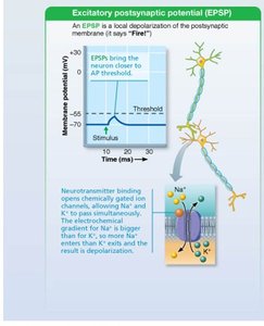
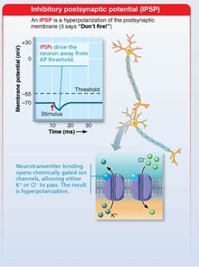
Neurotransmitter receptors on the postsynaptic membrane cause graded potentials, whose strength depends on the amount and duration of neurotransmitter in the cleft. There are two main types:
EPSP (Excitatory Postsynaptic Potential): Depolarizes the postsynaptic membrane, bringing it closer to threshold for action potential generation.
IPSP (Inhibitory Postsynaptic Potential): Hyperpolarizes the postsynaptic membrane, moving it further from threshold and inhibiting action potential generation.
Summation of Postsynaptic Potentials
Integration and Modification
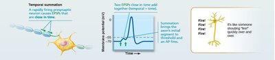
A single EPSP is usually insufficient to trigger an action potential. However, EPSPs can summate to influence the postsynaptic neuron. Summation can be:
Temporal summation: Multiple impulses from one presynaptic neuron in rapid succession add together.
Spatial summation: Simultaneous stimulation by multiple presynaptic neurons adds together.
Synaptic Potentiation
Mechanisms and Importance
Repeated use of a synapse increases the ability of the presynaptic cell to excite the postsynaptic neuron, a process called synaptic potentiation. This is associated with increased Ca2+ in the presynaptic terminal, leading to more neurotransmitter release and more EPSPs. Long-term potentiation is important for learning and memory.
Neurotransmitters
Classification and Examples
Neurotransmitters are the chemical messengers of the nervous system. Over 50 have been identified, and most neurons produce more than one. They are classified by chemical structure and function:
Class | Examples | Notes |
|---|---|---|
Acetylcholine (ACh) | Neuromuscular junctions, ANS, CNS | First identified, best understood |
Biogenic amines | Dopamine, norepinephrine, epinephrine, serotonin, histamine | Roles in emotion, biological clock; imbalances linked to mental illness |
Peptides | Substance P, endorphins, somatostatin, cholecystokinin | Pain modulation, digestion regulation |
Purines | ATP, adenosine | ATP as neurotransmitter; adenosine is inhibitory |
Gases & lipids | Nitric oxide (NO), carbon monoxide (CO), hydrogen sulfide (H2S) | Involved in memory, smooth muscle relaxation |
Endocannabinoids | Act at THC receptors | Learning, memory, appetite, nausea suppression |
Neurotransmitter Effects and Actions
Excitatory vs. Inhibitory Effects
Effect depends on the receptor type, not just the neurotransmitter.
GABA and glycine are usually inhibitory; glutamate is usually excitatory.
Acetylcholine can be excitatory (skeletal muscle) or inhibitory (cardiac muscle).
Direct vs. Indirect Actions
Direct action: Neurotransmitter binds directly to and opens ion channels (e.g., ACh, amino acids) for rapid responses.
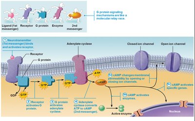
Indirect action: Neurotransmitter acts through second messengers (e.g., G protein pathways), producing broader, longer-lasting effects (e.g., biogenic amines, neuropeptides, gases).
Neurotransmitter Receptors
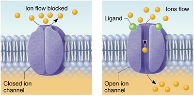
Channel-Linked Receptors
These are ligand-gated ion channels that mediate immediate, brief synaptic transmission. Excitatory receptors allow Na+ influx (depolarization), while inhibitory receptors allow Cl– influx (hyperpolarization).
G Protein–Linked Receptors
These receptors produce indirect, complex, slow, and often prolonged responses via second messengers such as cyclic AMP (cAMP). They are involved in widespread metabolic changes and gene activation.
Neural Integration and Circuits
Neuronal Pools and Processing
Neurons function in groups called neuronal pools, integrating incoming information and forwarding processed information to other destinations. Neural integration allows for complex behaviors and responses.
Patterns of Neural Processing
Parallel processing: Input travels along several pathways, allowing for simultaneous processing and multiple responses (e.g., a smell triggering memories and emotions).
Types of Circuits in Neuronal Pools
Type | Description | Example |
|---|---|---|
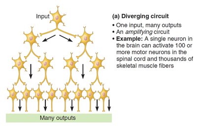
Diverging circuit | One input, many outputs; amplifies signals | Single neuron activating many motor units |
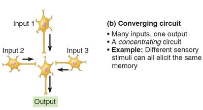
Converging circuit | Many inputs, one output; concentrates signals | Different sensory stimuli eliciting the same memory |
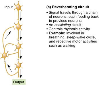
Reverberating circuit | Signal travels through a chain, feeding back to earlier neurons; oscillates | Breathing, sleep-wake cycle, walking |
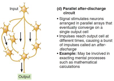
Parallel after-discharge circuit | Signal stimulates neurons in parallel arrays converging on one output | Mental processes like calculations |
References
Marieb, E. N., & Hoehn, K. (2023). Human Anatomy and Physiology (12th ed.). Pearson.
