 Back
BackFundamentals of the Nervous System and Nervous Tissue: Synapses, Neurotransmitters, and Neural Integration
Study Guide - Smart Notes
 Tailored notes based on your materials, expanded with key definitions, examples, and context.
Synapses: Structure and Function
Definition and Types of Synapses
Synapses are specialized junctions that mediate information transfer between neurons or between a neuron and an effector cell. They are essential for the functioning of the nervous system, allowing communication and integration of signals.
Presynaptic neuron: Conducts impulses toward the synapse and sends information.
Postsynaptic neuron: Transmits electrical signals away from the synapse and receives information. In the peripheral nervous system (PNS), the postsynaptic cell may be a neuron, muscle cell, or gland cell.
Types of Synaptic Connections
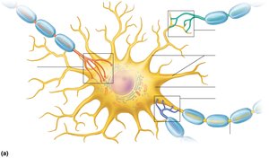
Axodendritic: Between axon terminals of one neuron and dendrites of another.
Axosomatic: Between axon terminals of one neuron and the soma (cell body) of another.
Axoaxonal: Between axon terminals of two neurons (less common).
Dendrodendritic: Between dendrites of two neurons (less common).
Somatodendritic: Between soma and dendrite (less common).
Chemical vs. Electrical Synapses
Chemical synapses: Most common; use neurotransmitters for signal transmission.
Electrical synapses: Less common; neurons are electrically coupled via gap junctions, allowing rapid, bidirectional communication. Found in some brain regions and are most abundant in embryonic nervous tissue.
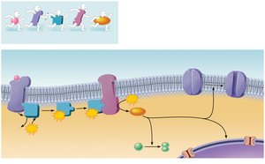
Chemical Synapses: Mechanism and Transmission
Structure and Function
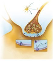
Chemical synapses are specialized for the release and reception of chemical neurotransmitters. They consist of:
Axon terminal: Contains synaptic vesicles filled with neurotransmitter.
Receptor region: Located on the postsynaptic neuron's membrane, usually on a dendrite or cell body.
Synaptic cleft: Fluid-filled space separating the two parts.
Steps in Chemical Synaptic Transmission
An action potential arrives at the axon terminal of the presynaptic neuron.
Voltage-gated Ca2+ channels open, allowing Ca2+ to enter the axon terminal.
Ca2+ entry causes synaptic vesicles to release neurotransmitter by exocytosis.
Neurotransmitter diffuses across the synaptic cleft and binds to specific receptors on the postsynaptic membrane.
Binding of neurotransmitter opens ion channels, resulting in graded potentials (excitatory or inhibitory).
Neurotransmitter effects are terminated by reuptake, enzymatic degradation, or diffusion away from the synapse.
Synaptic Delay
The time required for neurotransmitter release, diffusion, and receptor binding (0.3 to 5.0 ms).
Synaptic delay is the rate-limiting step of neural transmission.
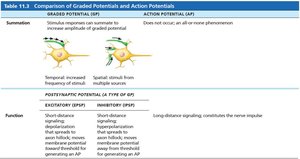
Postsynaptic Potentials: EPSPs and IPSPs
Types of Postsynaptic Potentials
Neurotransmitter receptors cause graded potentials that vary in strength based on the amount and duration of neurotransmitter release. There are two main types:
c (EPSPs): Depolarize the postsynaptic membrane, increasing the likelihood of action potential generation.
Inhibitory postsynaptic potentials (IPSPs): Hyperpolarize the postsynaptic membrane, decreasing the likelihood of action potential generation.
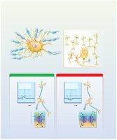
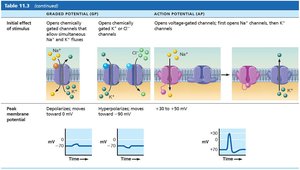
Mechanism of EPSPs
Neurotransmitter binding opens chemically gated channels for Na+ and K+.
Na+ influx is greater than K+ efflux, resulting in depolarization.
If the EPSP reaches threshold, it triggers an action potential at the axon hillock.
Mechanism of IPSPs
Neurotransmitter binding opens channels for K+ (out) or Cl– (in), causing hyperpolarization.
Moves the membrane potential farther from threshold, inhibiting action potential generation.
Summation of Postsynaptic Potentials
Temporal summation: Rapid, successive impulses from one presynaptic neuron add together.
Spatial summation: Simultaneous stimulation by multiple presynaptic neurons adds together.
Synaptic Potentiation and Presynaptic Inhibition
Synaptic potentiation: Repeated use of a synapse increases its ability to excite the postsynaptic neuron, often involving increased Ca2+ concentration and kinase activation.
Presynaptic inhibition: Release of excitatory neurotransmitter is inhibited by another neuron, reducing EPSPs.
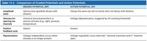
Comparison of Graded Potentials and Action Potentials
Key Differences
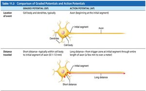
Graded potentials and action potentials are two types of electrical signals in neurons, differing in location, amplitude, and function.
Feature | Graded Potential (GP) | Action Potential (AP) |
|---|---|---|
Location | Cell body and dendrites | Axon (initial segment) |
Distance traveled | Short (1-2 mm) | Long (up to a meter) |
Feature | Graded Potential (GP) | Action Potential (AP) |
|---|---|---|
Amplitude | Various sizes, decays with distance | Always same size, does not decay |
Stimulus | Chemical (neurotransmitter), mechanical, or temperature | Voltage depolarization (threshold) |
Positive feedback | Absent | Present |
Repolarization | Voltage-independent | Voltage-regulated |
Feature | Graded Potential (GP) | Action Potential (AP) |
|---|---|---|
Summation | Can summate (temporal/spatial) | Does not summate |
Function | EPSP/IPSP: short-distance signaling | Long-distance signaling |
Feature | Graded Potential (GP) | Action Potential (AP) |
|---|---|---|
Initial effect | Opens chemically gated channels | Opens voltage-gated channels |
Peak membrane potential | Depolarizes/hyperpolarizes | +30 to +50 mV |
Neurotransmitters: Classification and Function
Overview
Neurotransmitters are the chemical language of the nervous system. Over 50 have been identified, and most neurons produce multiple types, exerting diverse influences depending on stimulation frequency and receptor type.
Classification by Chemical Structure
Acetylcholine (ACh): First identified; released at neuromuscular junctions, some CNS and ANS neurons. Synthesized by choline acetyltransferase, degraded by acetylcholinesterase.
Biogenic amines:
Catecholamines: Dopamine, norepinephrine (NE), epinephrine (from tyrosine).
Indolamines: Serotonin (from tryptophan), histamine (from histidine).
Amino acids: Glutamate, aspartate, glycine, GABA (gamma-aminobutyric acid).
Peptides (neuropeptides): Substance P (pain), endorphins (natural opiates), gut-brain peptides (somatostatin, cholecystokinin).
Purines: ATP (energy molecule, now considered a neurotransmitter), adenosine (inhibitor in brain).
Gases and lipids: Nitric oxide (NO), carbon monoxide (CO), hydrogen sulfide (H2S); lipid-soluble, synthesized on demand.
Endocannabinoids: Act at same receptors as THC; involved in learning, memory, appetite, and nausea suppression.
Classification by Function
Effects:
Excitatory: Depolarizing (e.g., glutamate).
Inhibitory: Hyperpolarizing (e.g., GABA, glycine).
Effect depends on receptor type (e.g., ACh is excitatory at skeletal muscle, inhibitory at cardiac muscle).
Actions:
Direct: Binds directly to and opens ion channels (rapid response; e.g., ACh, amino acids).
Indirect: Acts through second messengers (G protein pathways; longer-lasting effects; e.g., biogenic amines, neuropeptides, gases).
Neuromodulator: Chemical messenger that modulates synaptic transmission strength without directly causing EPSPs or IPSPs.
Neurotransmitter Receptors
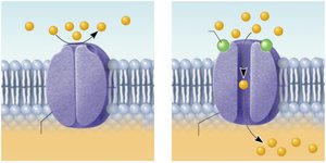
Channel-Linked Receptors
Ligand-gated ion channels that mediate rapid, brief synaptic transmission. Excitatory receptors allow Na+ influx (depolarization), while inhibitory receptors allow Cl– influx (hyperpolarization).
G Protein–Linked Receptors
These receptors mediate indirect, complex, and prolonged responses via second messengers (e.g., cAMP, Ca2+). They can open/close ion channels, activate enzymes, or induce gene expression.
Neural Integration: Neuronal Pools and Processing
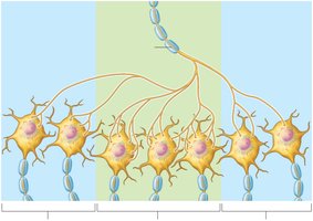
Neuronal Pools
Neuronal pools are functional groups of neurons that integrate incoming information and forward processed information to other destinations. They consist of:
Discharge zone: Neurons closest to the incoming fiber, most likely to generate an impulse.
Facilitated zone: Neurons farther away, usually not excited to threshold unless stimulated by another source.
Patterns of Neural Processing
Serial processing: Input travels along one pathway to a specific destination; produces specific, anticipated responses (e.g., spinal reflex arc).
Parallel processing: Input travels along several pathways; promotes numerous responses and is important for higher-level mental functioning.
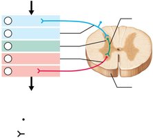
Reflex Arcs
Reflexes are rapid, automatic responses to stimuli, occurring over pathways called reflex arcs with five components:
Receptor
Sensory neuron
CNS integration center
Motor neuron
Effector
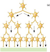
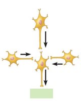
Types of Circuits in Neuronal Pools
Diverging circuit: One input, many outputs; amplifies signals (e.g., one neuron activates many motor neurons).
Converging circuit: Many inputs, one output; concentrates signals (e.g., different sensory stimuli elicit the same memory).
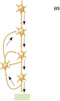
Reverberating circuit: Signal travels through a chain of neurons, each feeding back to previous neurons; controls rhythmic activities (e.g., breathing).
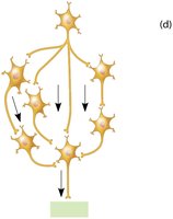
Parallel after-discharge circuit: Signal stimulates neurons arranged in parallel arrays that converge on a single output cell; may be involved in complex mental processes.
Summary Table: Comparison of Graded Potentials and Action Potentials
Feature | Graded Potential (GP) | Action Potential (AP) |
|---|---|---|
Location | Cell body, dendrites | Axon (initial segment) |
Distance | Short | Long |
Amplitude | Variable, decays | Constant, does not decay |
Stimulus | Chemical, mechanical, temperature | Voltage (threshold) |
Summation | Possible | Not possible |
Function | EPSP/IPSP, short-distance | Long-distance signaling |
Key Equations
Membrane potential change (graded potential): where is the change in membrane potential, is the current, and is the resistance.
Action potential threshold:
Additional info: Tables and diagrams have been expanded and described for clarity. All images included are directly relevant to the adjacent content and reinforce key concepts in synaptic transmission, neural integration, and circuit types.
