 Back
BackFundamentals of the Nervous System and Nervous Tissue: Structured Study Notes
Study Guide - Smart Notes
 Tailored notes based on your materials, expanded with key definitions, examples, and context.
Fundamentals of the Nervous System and Nervous Tissue
The Nervous System: Overview and Functions
The nervous system is the master controlling and communicating system of the body. It uses electrical and chemical signals to rapidly and specifically coordinate bodily functions, often resulting in immediate responses.
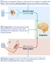
Sensory Input: Information is gathered by sensory receptors about internal and external changes. Sensory input travels via afferent pathways to the central nervous system (CNS).
Integration: The CNS processes and interprets sensory input, deciding on an appropriate response.
Motor Output: Activation of effectors (muscles and glands) produces a response via efferent pathways.
Organization of the Nervous System
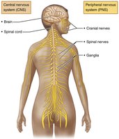
The nervous system is divided into two principal parts: the central nervous system (CNS) and the peripheral nervous system (PNS).
CNS: Consists of the brain and spinal cord, serving as the integration and control center.
PNS: Composed of nerves and receptors outside the CNS, including cranial and spinal nerves, and the enteric nervous system in the gastrointestinal tract.
Functional Divisions of the PNS
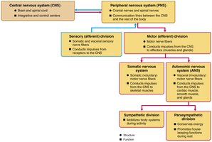
Sensory (Afferent) Division: Somatic sensory fibers convey impulses from skin, skeletal muscles, and joints; visceral sensory fibers convey impulses from visceral organs.
Motor (Efferent) Division: Transmits impulses from CNS to effectors. Includes the somatic (voluntary) nervous system and the autonomic (involuntary) nervous system.
Autonomic Nervous System (ANS): Regulates smooth muscle, cardiac muscle, and glands. Subdivided into sympathetic (mobilizes body systems) and parasympathetic (conserves energy, promotes housekeeping functions).
Neuroglia and Neurons
Neuroglia (Glial Cells)
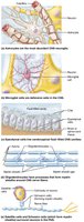
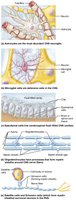
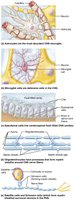
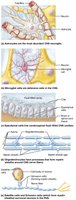
Neuroglia are supporting cells in nervous tissue, surrounding and wrapping delicate neurons. They are essential for neuron function and maintenance.
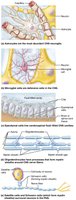
Astrocytes: Most abundant glial cells in CNS; support neurons, regulate exchanges, guide migration, control chemical environment, and recycle neurotransmitters.
Microglial Cells: Monitor neuron health, migrate toward injured neurons, and phagocytize debris.
Ependymal Cells: Line CNS cavities, circulate cerebrospinal fluid (CSF), and form a barrier between CSF and tissue fluid.
Oligodendrocytes: Form myelin sheaths in CNS, insulating axons.
Satellite Cells (PNS): Surround neuron cell bodies, similar function to astrocytes.
Schwann Cells (PNS): Form myelin sheaths around peripheral nerve fibers, vital for regeneration.
Neurons: Structure and Function
Neurons are excitable cells that transmit electrical signals. They exhibit extreme longevity, are mostly amitotic, and have a high metabolic rate.
Cell Body (Neurosoma): Contains nucleus, nucleolus, and cytoplasm; biosynthetic and metabolic center.
Processes: Dendrites (receptive/input regions) and axons (conducting/output regions).
Nuclei: Clusters of neuron cell bodies in CNS.
Ganglia: Clusters of neuron cell bodies in PNS.
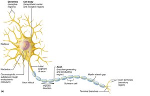
Dendrites
Short, branched processes; receive signals and convey them toward the cell body as graded potentials.
Contain dendritic spines for specialized information collection.
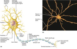
Axons
Single, long process; generates and transmits nerve impulses away from cell body.
Axon terminals release neurotransmitters.
Bundles in CNS are called tracts; in PNS, nerves.
Axonal transport: anterograde (away from cell body) and retrograde (toward cell body).
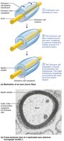
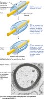
Myelin Sheath
White, fatty substance insulating axons; increases speed of impulse transmission.
Formed by Schwann cells in PNS and oligodendrocytes in CNS.
Myelin sheath gaps (nodes of Ranvier) are sites for axon collateral emergence.
Classification of Neurons
Structural Classification
Neurons are classified by the number of processes extending from the cell body.
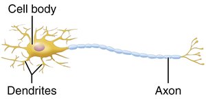
Multipolar: Many processes (1 axon, many dendrites); most common in CNS.
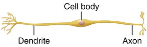
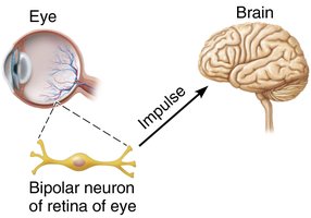
Bipolar: Two processes (1 axon, 1 dendrite); rare, found in retina, ear, olfactory mucosa.
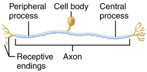
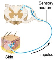
Unipolar: One process (T-shaped); found mainly in PNS ganglia, function as sensory neurons.
Neuron Type | Processes | Location |
|---|---|---|
Multipolar | 1 axon, many dendrites | CNS |
Bipolar | 1 axon, 1 dendrite | Retina, ear, olfactory mucosa |
Unipolar | 1 process (T-shaped) | PNS ganglia |
Functional Classification
Sensory (Afferent) Neurons: Transmit impulses toward CNS; mostly unipolar.
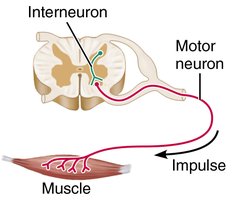
Motor (Efferent) Neurons: Carry impulses from CNS to effectors; multipolar.
Interneurons: Shuttle signals within CNS; most abundant.
Functional Class | Direction | Structure |
|---|---|---|
Sensory | Toward CNS | Unipolar |
Motor | From CNS to effectors | Multipolar |
Interneuron | Within CNS | Multipolar |
Membrane Potentials and Electrical Properties
Basic Principles of Electricity
Neurons have a resting membrane potential and can rapidly change it, making them highly excitable.
Voltage: Potential energy generated by separated charge.
Current: Flow of electrical charge (ions).
Resistance: Hindrance to charge flow.
Ohm's Law:
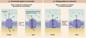
Ion Channels
Leakage Channels: Always open.
Gated Channels: Open/close in response to stimuli (chemical, voltage, mechanical).
Resting Membrane Potential
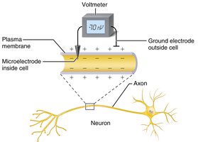
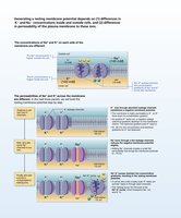
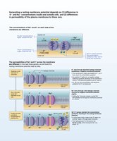
Resting membrane potential is typically -70 mV, with the inside of the cell more negative than the outside. Generated by differences in ionic composition and membrane permeability.
High K+ inside, high Na+ outside.
Membrane more permeable to K+ than Na+.
Sodium-potassium pump maintains gradients: 3 Na+ out, 2 K+ in.
Changes in Membrane Potential
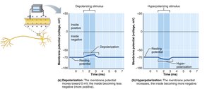
Depolarization: Membrane potential moves toward zero; increases probability of impulse.
Hyperpolarization: Membrane potential moves away from zero; decreases probability of impulse.
Graded Potentials and Action Potentials
Graded Potentials
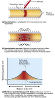
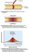
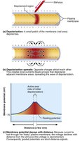
Graded potentials are short-lived, localized changes in membrane potential, essential for initiating action potentials.
Triggered by opening of gated ion channels.
Spread and decay quickly; act as signals over short distances.
Action Potentials
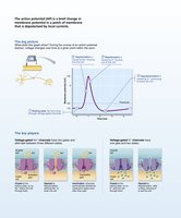
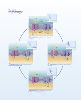
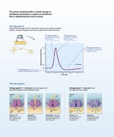
An action potential (AP) is a brief reversal of membrane potential, typically from -70 mV to +30 mV. APs are long-distance signals generated in axons and do not decay over distance.
Four Main Steps: Resting state, depolarization, repolarization, hyperpolarization.
Voltage-gated Na+ and K+ channels play key roles.
All-or-none phenomenon: AP either happens completely or not at all.
Propagation and Coding
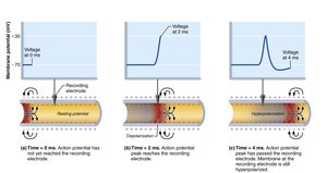
APs propagate along axons, self-propagating in a forward direction.
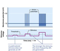
Stimulus intensity is coded by frequency of APs, not amplitude.
Refractory Periods
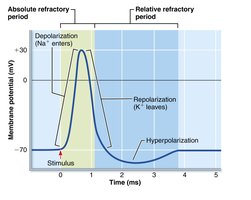
Absolute Refractory Period: No new AP can be generated.
Relative Refractory Period: AP can be generated only by a strong stimulus.
Conduction Velocity
Depends on axon diameter and degree of myelination.
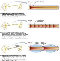
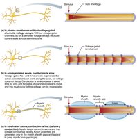
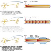
Continuous conduction in nonmyelinated axons; saltatory conduction in myelinated axons (much faster).
Synapses and Neural Communication
Synapses
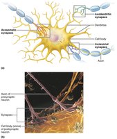
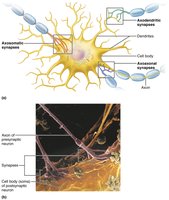
Synapses are junctions that mediate information transfer between neurons or between a neuron and an effector cell.
Presynaptic Neuron: Sends information.
Postsynaptic Neuron: Receives information.
Types: axodendritic, axosomatic, axoaxonal, dendrodendritic, somatodendritic.
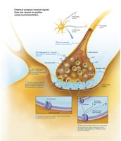
Two main types: electrical (gap junctions, rapid) and chemical (neurotransmitter-mediated).
Chemical Synapses
Most common; specialized for release and reception of neurotransmitters.
Six steps: AP arrives, Ca2+ enters, neurotransmitter released, diffuses, binds to receptor, ion channels open, effect terminated.
Postsynaptic Potentials and Integration
Postsynaptic Potentials
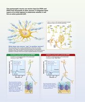
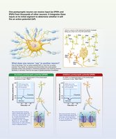
EPSP (Excitatory): Depolarizes postsynaptic membrane, increases likelihood of AP.
IPSP (Inhibitory): Hyperpolarizes postsynaptic membrane, decreases likelihood of AP.
Summation
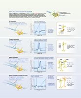
Temporal Summation: Rapid-fire impulses from one neuron.
Spatial Summation: Simultaneous stimulation by multiple neurons.
Neurotransmitters
Classification by Chemical Structure
Acetylcholine: Released at neuromuscular junctions, degraded by acetylcholinesterase.
Biogenic Amines: Dopamine, norepinephrine, epinephrine, serotonin, histamine.
Amino Acids: Glutamate, aspartate, glycine, GABA.
Peptides: Substance P, endorphins, somatostatin, CCK.
Purines: ATP, adenosine.
Gases and Lipids: Nitric oxide, carbon monoxide, hydrogen sulfide, endocannabinoids.
Classification by Function
Effects: Excitatory (depolarizing) or inhibitory (hyperpolarizing), depending on receptor.
Actions: Direct (fast, opens ion channels) or indirect (slow, via second messengers).
Neuromodulators: Affect strength of synaptic transmission, act locally.
Neurotransmitter Receptors
Channel-Linked Receptors: Ligand-gated ion channels, immediate and brief action.
G Protein–Linked Receptors: Indirect, complex, slow, prolonged responses via second messengers.
Neural Integration and Circuits
Patterns of Neural Processing
Serial Processing: Input travels along one pathway; produces specific response (e.g., reflex arc).
Parallel Processing: Input travels along several pathways; promotes higher-level functions.
Types of Neural Circuits
Diverging: One input, many outputs.
Converging: Many inputs, one output.
Reverberating: Repetitive signals.
Parallel After-Discharge: Multiple pathways, single output.
Developmental Aspects of Neurons
Neural Development
Nervous system originates from neural tube and neural crest (ectoderm).
Neuroblasts migrate, connect, and differentiate into neurons.
Growth cones guide axons to targets using adhesion proteins and neurotropins.
Astrocytes support synapse formation; apoptosis eliminates unused neurons.
Learning reinforces synapses; excessive pruning may relate to schizophrenia.
Some neurons (olfactory, hippocampus) continue to divide after birth.
Example: Nerve growth factor (NGF) keeps neuroblasts alive; filopodia follow signals to target.
Additional info: These notes provide a comprehensive overview of Chapter 11, suitable for exam preparation in an introductory anatomy and physiology course.
