 Back
BackFundamentals of the Nervous System: Structure and Function
Study Guide - Smart Notes
 Tailored notes based on your materials, expanded with key definitions, examples, and context.
Fundamentals of the Nervous System
Overview and Functions
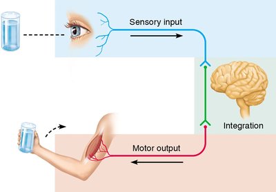
The nervous system is the master controlling and communicating system of the body. It is responsible for regulating and coordinating body activities through rapid transmission of electrical signals. The nervous system has three main, overlapping functions:
Sensory input: Gathering information from sensory receptors about internal and external changes.
Integration: Processing and interpreting sensory input to determine an appropriate response.
Motor output: Activating effector organs (muscles and glands) to cause a response.
Organization of the Nervous System
Central and Peripheral Nervous Systems
The nervous system is divided into two principal parts:
Central Nervous System (CNS): Consists of the brain and spinal cord; responsible for integration and command.
Peripheral Nervous System (PNS): Consists of nerves and ganglia outside the CNS; connects the CNS to limbs and organs.
Functional Subdivisions of the PNS
Sensory (afferent) division: Transmits sensory information to the CNS.
Somatic sensory fibers: Convey impulses from skin, skeletal muscles, and joints.
Visceral sensory fibers: Transmit impulses from visceral organs.
Motor (efferent) division: Transmits commands from the CNS to effector organs.
Somatic nervous system: Controls voluntary movements of skeletal muscles.
Autonomic nervous system (ANS): Regulates involuntary functions (e.g., heart, glands).
Sympathetic division: Mobilizes body systems during activity ("fight or flight").
Parasympathetic division: Conserves energy and promotes "rest and digest" functions.
Nervous Tissue
Principal Cell Types
Nervous tissue is composed of two main cell types:
Neuroglia (glial cells): Support, protect, and insulate neurons.
Neurons (nerve cells): Excitable cells that transmit electrical signals.
Types of Neuroglia
CNS Neuroglia:
Astrocytes: Support and brace neurons, regulate the environment.
Microglial cells: Act as phagocytes, removing debris and pathogens.
Ependymal cells: Line cerebrospinal fluid-filled cavities, help circulate CSF.
Oligodendrocytes: Produce myelin sheaths in the CNS.
PNS Neuroglia:
Satellite cells: Surround neuron cell bodies in the PNS.
Schwann cells: Form myelin sheaths in the PNS.
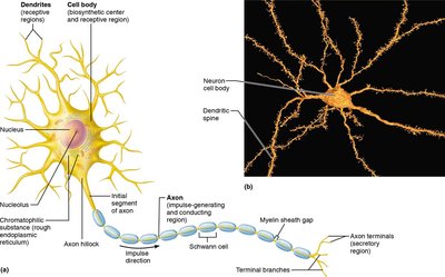
Neurons: Structure and Function
Neurons are the structural and functional units of the nervous system. They possess:
Extreme longevity: Can function for a lifetime.
Amitotic: Most do not divide after development.
High metabolic rate: Require continuous supply of oxygen and glucose.
All neurons have:
Cell body (soma or perikaryon): Contains the nucleus and organelles; clusters in CNS are called nuclei, in PNS ganglia.
Processes: Extensions from the cell body; bundles in CNS are tracts, in PNS nerves.
There are two main types of processes:
Dendrites: Receive incoming signals.
Axons: Transmit impulses away from the cell body.
Classification of Neurons and Nerves
Functional classification of neurons:
Sensory neurons: Transmit impulses toward the CNS.
Motor neurons: Carry impulses away from the CNS to effectors.
Interneurons: Connect sensory and motor neurons within the CNS.
Nerves are classified by impulse direction:
Sensory nerves: Carry impulses toward the CNS.
Motor nerves: Carry impulses away from the CNS.
Mixed nerves: Contain both sensory and motor fibers.
Nerve Impulse Physiology
Resting Membrane Potential and Ion Channels
Neurons maintain a resting membrane potential, meaning they are polarized (inside is negative relative to outside). Voltage is the potential energy measured between two points. Plasma membranes contain selective ion channels:
Leakage channels: Always open, allow ions to move down their gradients.
Gated channels: Open or close in response to stimuli.
Chemically gated
Voltage-gated
Mechanically gated
Membrane Potential Changes and Signals
Changes in ion concentration or membrane permeability can cause:
Depolarization: Inside becomes less negative.
Hyperpolarization: Inside becomes more negative.
These changes produce two types of signals:
Graded potentials: Short-distance signals.
Action potentials: Long-distance signals of axons.
Generation of an Action Potential
Resting state
Depolarization
Repolarization
Hyperpolarization
During the refractory period, the neuron cannot fire another action potential. There are two types:
Absolute refractory period
Relative refractory period
Synapses and Neurotransmitters
A synapse is a functional junction between two neurons or between a neuron and an effector cell. Types include:
Electrical synapse
Chemical synapse
Neurotransmitters are classified by:
Molecular structure
Function: Excitatory or inhibitory effects; direct or indirect actions
Central Nervous System (CNS)
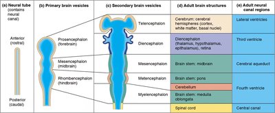
Embryonic Development of the Brain and Spinal Cord
The CNS develops from the embryonic neural tube, which forms three primary brain vesicles:
Prosencephalon (forebrain)
Mesencephalon (midbrain)
Rhombencephalon (hindbrain)
These give rise to five secondary brain vesicles, which develop into the adult brain structures and associated ventricles.
Major Regions of the Brain
Cerebrum: Includes the cerebral cortex (motor, sensory, and association areas), white matter, and basal nuclei. Surface features include gyri, sulci, and fissures.
Diencephalon: Contains the thalamus, hypothalamus (vital for homeostasis, includes mammillary bodies and infundibulum), pituitary gland, and epithalamus (pineal gland).
Brain Stem: Composed of the midbrain (cerebral peduncles, corpora quadrigemina), pons, and medulla oblongata.
Cerebellum: Coordinates voluntary movements and balance.
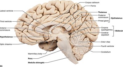
Protection of the Brain
Meninges: Three connective tissue layers—dura mater, arachnoid mater, pia mater.
Cerebrospinal fluid (CSF): Cushions and nourishes the brain.
Blood Brain Barrier: Protects the brain from harmful substances in the blood.
Spinal Cord
The spinal cord is a major CNS structure responsible for transmitting signals and mediating reflexes.
Peripheral Nervous System (PNS)
Sensation and Perception
Survival depends on the ability to sense and perceive environmental changes. Sensory receptors are classified by:
Stimulus type:
Mechanoreceptors
Thermoreceptors
Photoreceptors
Chemoreceptors
Nociceptors
Location:
Exteroreceptors
Interoceptors
Proprioceptors
