Back
Unit 4- Ch 19 Pt 2 Gas Exchange, Transport, and Control of Ventilation
Study Guide - Smart Notes
 Tailored notes based on your materials, expanded with key definitions, examples, and context.
Gas Transport in Blood
Carbon Dioxide Transport and Buffering
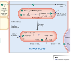
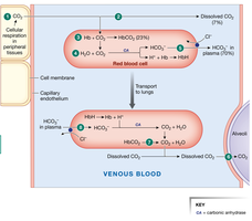
Carbon dioxide (CO2) produced by cellular respiration in systemic tissues is transported in the blood to the lungs for exhalation. CO2 is carried in three main forms:
Dissolved CO2: About 7% of total CO2 is transported dissolved directly in plasma, proportional to its partial pressure (PCO2).
Carbaminohemoglobin (HbCO2): Approximately 23% of CO2 binds to deoxyhemoglobin, forming carbaminohemoglobin.
Bicarbonate Ion (HCO3-): The majority (~70%) of CO2 is converted to bicarbonate in red blood cells (RBCs) via the enzyme carbonic anhydrase (CA):
Chloride Shift: To maintain electrical neutrality, HCO3- exits RBCs in exchange for Cl- ions (chloride shift).
Buffering: H+ ions produced are buffered by hemoglobin (Hb), forming HHb.
Gas Exchange in the Lungs
In the lungs, the processes reverse to allow CO2 to diffuse into the alveoli for exhalation:
Bicarbonate re-enters RBCs (reverse chloride shift), recombines with H+ to form CO2 and H2O (via CA).
CO2 dissociates from HbCO2 and diffuses into the alveolus.
Dissolved CO2 in plasma also diffuses into the alveolus.
Carbon Monoxide and Hemoglobin
Carbon monoxide (CO) binds to hemoglobin with 210 times the affinity of oxygen (O2), occupying the O2 binding site and forming carboxyhemoglobin. This drastically reduces O2 delivery to tissues (anemic hypoxia) and causes a leftward shift in the hemoglobin dissociation curve. CO poisoning does not trigger the ventilation reflex because arterial PO2 does not decrease, leading to potential asphyxiation.
Symptoms of CO poisoning: Headache, dizziness, weakness, nausea, confusion, and potentially loss of consciousness or death.
Control of Ventilation
Neural Regulation of Breathing
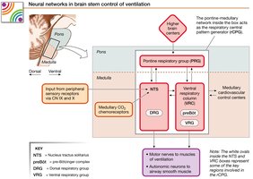
Ventilation is regulated by neural networks in the brainstem, integrating sensory input and coordinating respiratory muscle activity.
Central Pattern Generator (rCPG): Located in the medulla and pons, generates rhythmic breathing patterns.
Pontine Respiratory Group (PRG): Integrates sensory information and modulates medullary centers.
Medullary Respiratory Centers:
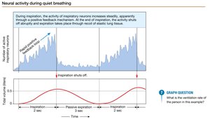
Dorsal Respiratory Group (DRG): Controls inspiratory muscles, exhibits a ramping pattern for inspiration (positive feedback), and ends stimulation for expiration.
Ventral Respiratory Group (VRG): Controls forced ventilation and accessory muscles (pharynx, larynx, tongue). The Pre-Bötzinger complex within the VRG may act as a pacemaker for respiratory rhythm.
Chemoreceptor Regulation
Chemoreceptors monitor blood gases and pH to adjust ventilation:
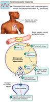
Peripheral Chemoreceptors: Located in carotid and aortic bodies, respond to changes in PO2, PCO2, and pH.
Activated by PO2 ≤ 60 mm Hg (hypoxia), increased H+ (metabolic acidosis), or increased PCO2 (respiratory acidosis).
Glomus cells are the primary receptor cells.
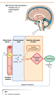
Central Chemoreceptors: Located in the medulla, respond to changes in PCO2 of cerebrospinal fluid (CSF) via the reaction:
Increased [H+] in CSF increases firing and stimulates ventilation.
Chemoreceptor Redundancy and Adaptation
Central and peripheral chemoreceptors interact to maintain homeostasis. In chronic lung disorders (chronic hypercapnia), the PCO2 setpoint is raised, making PO2 more important for stimulating ventilation. Hyperventilation can reduce PCO2 below the threshold for stimulation, decreasing alveolar ventilation and increasing systemic arterial PCO2.
Protective Reflexes
Protective reflexes prevent aspiration and injury:
Cough Reflex: Triggered by receptors in the larynx, trachea, and bronchi; involves deep inspiration and forceful expiration to clear lower airways.
Sneeze Reflex: Triggered by receptors in the nasal cavity and pharynx; clears upper airways.
Irritant Receptors: Detect noxious chemicals, causing bronchoconstriction and cessation of breathing.
Hering-Breuer Inflation Reflex: Lung stretch receptors prevent over-inflation, especially important in infants.
Higher Center Modulation of Breathing
Higher brain centers (hypothalamus, cerebrum, limbic system) can voluntarily override or modulate breathing for activities such as speech, swallowing, swimming, and voluntary apnea. Hyperventilation before breath-holding can lower PCO2, delaying the urge to breathe but risking hypoxia and unconsciousness.
Summary Tables
Major Forms of CO2 Transport in Blood
Form | Percentage | Description |
|---|---|---|
Dissolved CO2 | ~7% | Directly dissolved in plasma |
Carbaminohemoglobin (HbCO2) | ~23% | Bound to hemoglobin |
Bicarbonate (HCO3-) | ~70% | Converted in RBCs, transported in plasma |
Key Respiratory Centers and Functions
Center | Location | Function |
|---|---|---|
Pontine Respiratory Group (PRG) | Pons | Modulates medullary centers, integrates sensory input |
Dorsal Respiratory Group (DRG) | Medulla | Controls inspiration, ramping pattern |
Ventral Respiratory Group (VRG) | Medulla | Controls forced ventilation, accessory muscles |
Pre-Bötzinger Complex | Medulla (VRG) | Possible pacemaker for respiratory rhythm |
Summary Diagrams
Use the provided summary figures as learning aids to reinforce the pathways of gas transport and the neural control of ventilation.