 Back
BackGeneral Sensation and Special Senses: Hearing and Equilibrium
Study Guide - Smart Notes
 Tailored notes based on your materials, expanded with key definitions, examples, and context.
General Sensation: Sensory Receptors and Neurons
Types of Sensory Receptors by Location
Sensory neurons are distributed throughout the body and respond to environmental changes. Each sensory neuron is specialized for a particular type of stimulus. The main types of sensory receptors are classified by their location:
Exteroceptors: Detect stimuli from outside the body, such as skin receptors and special sense organs.
Interoceptors: Detect stimuli from within the body, including organs and smooth muscle.
Proprioceptors: Detect internal stimuli related to the skeletal system, such as stretch sensors in tendons.
Functional Components of Sensation
Sensory input to the brain is processed through two main components:
Sensation: Awareness of a stimulus.
Perception: Interpretation of the stimulus by the central nervous system (CNS).
Classification of Sensory Receptor Neurons
Sensory neurons can be classified in three ways:
Type of stimulus: Mechanoreceptors (touch, pressure, vibration, stretch), Thermoreceptors (temperature), Photoreceptors (light), Chemoreceptors (chemicals), Nociceptors (pain).
Body location: Exteroceptors, Interoceptors, Proprioceptors.
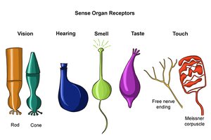
Structural complexity: Simple (general senses) and Special (special senses).
Structural Classification of Sensory Receptors
Simple Receptors: Nonencapsulated
Simple receptors are modified dendritic endings of sensory neurons and monitor most types of sensory information. Nonencapsulated simple receptors include:
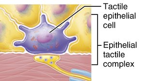
Tactile discs (Merkel discs): Stimulated by light touch.
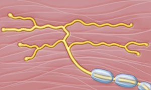
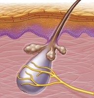
Hair follicle receptors: Stimulated by light touch from hair movement.
Simple Receptors: Encapsulated
Encapsulated simple receptors are surrounded by connective tissue and include:
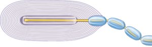
Tactile corpuscles (Meissner’s): Small, sensitive to discriminative touch, found in hairless areas.
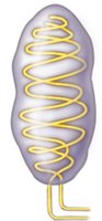
Lamellar corpuscles (Pacinian): Large, respond to deep pressure and vibration, located in deep dermis.
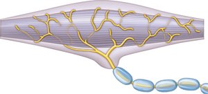
Bulbous corpuscles (Ruffini endings): Respond to deep, continuous pressure, found in the dermis.
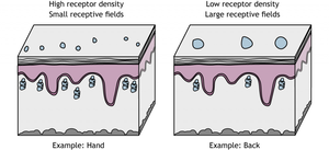
Receptor Density and Receptive Fields
Receptive Field Size and Receptor Density
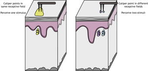
The ability to distinguish between stimuli depends on the density of sensory neurons and the size of their receptive fields. Areas with high receptor density and small receptive fields (e.g., fingertips) have greater sensory discrimination than areas with low density and large fields (e.g., back).
Special Senses: Hearing
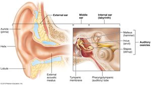
Anatomy of the Ear
The ear is divided into three major regions:
External ear: Captures and transfers sound to the middle ear.
Middle ear: Transfers and amplifies sound vibrations.
Internal ear: Responsible for hearing and equilibrium.

External Ear
Auricle (pinna): Elastic cartilage that collects sound.
External acoustic meatus: Tunnel leading to the tympanic membrane.
Tympanic membrane: Vibrates in response to sound waves and transmits vibrations to the ossicles.

Middle Ear
Tympanic cavity: Contains auditory ossicles (malleus, incus, stapes).
Oval window: Separates middle and inner ear, transmits vibrations to cochlear fluid.
Eustachian tube: Equalizes pressure in the middle ear.
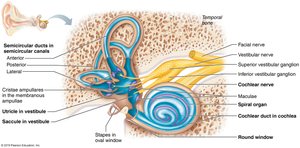
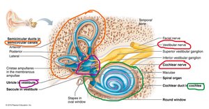
Inner Ear
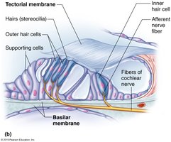
Cochlea: Contains the spiral organ and hair cells for hearing.
Hair cells: Specialized sensory receptors that respond to fluid vibrations.
Mechanism of Hearing
Sound waves are transmitted through the external and middle ear to the inner ear, where they cause vibrations in the cochlear fluid. These vibrations bend hair cells, which convert the physical stimulus into electrical signals. Different hair cells respond to different sound frequencies.
Special Senses: Equilibrium
Equilibrium Sensation
The inner ear also senses equilibrium through the semicircular canals and vestibule:
Semicircular canals: Detect rotational movement via fluid displacement.
Vestibule (utricle and saccule): Detect orientation relative to gravity.
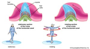
Mechanism of Equilibrium
Fluid movement in the semicircular canals and vestibule bends sensory cells, providing information about head movement and position. This mechanism is similar to sound conduction in the cochlea.
Clinical Tests for Hearing and Equilibrium
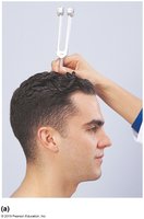
Weber Test
The Weber test checks for unilateral hearing loss by placing a tuning fork on the skull. It helps differentiate between conductive and sensorineural hearing loss.
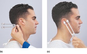
Rinne Test
The Rinne test compares air and bone conduction in each ear to further distinguish types of hearing loss.
Romberg Test
The Romberg test assesses equilibrium by observing swaying with eyes open or closed, indicating possible cerebellar or sensory system issues.
Summary Table: Types of Sensory Receptors
Type | Stimulus | Location | Example |
|---|---|---|---|
Exteroceptor | External | Skin, special sense organs | Touch, vision |
Interoceptor | Internal | Organs | Stretch in intestines |
Proprioceptor | Internal | Skeletal system | Tendon stretch |
Key Equations
Frequency of sound measured in cycles per second (Hz):
Where f is frequency and T is period.
Summary: Sensory receptors are specialized for different stimuli and locations, and their structure determines their function. The ear is a complex organ responsible for both hearing and equilibrium, with clinical tests available to assess function and diagnose disorders.
