 Back
BackGross Anatomy of the Muscular System: Structure, Function, and Organization
Study Guide - Smart Notes
 Tailored notes based on your materials, expanded with key definitions, examples, and context.
Gross Anatomy of the Muscular System
Muscle Actions and Interactions
The muscular system is responsible for producing movement by contracting and pulling on bones. Muscles work in coordinated groups to perform specific actions, with each muscle playing a distinct role in movement.
Prime Mover (Agonist): The main muscle responsible for a specific movement.
Antagonist: A muscle that opposes or reverses the action of the prime mover. Prime movers and antagonists are typically located on opposite sides of the joint they act upon.
Synergist: Assists the prime mover by adding extra force or reducing unnecessary movement.
Fixator: A type of synergist that stabilizes the origin of the prime mover.
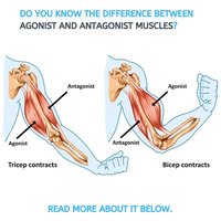
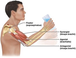
Example: During elbow flexion, the biceps brachii acts as the agonist, the triceps brachii as the antagonist, and the brachialis as a synergist.
Inferring Muscle Action by Position
The action of a muscle can often be predicted by its position relative to the joint it crosses:
Anterior side: Usually produces flexion (except at the knee and ankle).
Posterior side: Usually produces extension (except at the knee and ankle).
Lateral side: Produces abduction.
Medial side: Produces adduction.
Example: The deltoid (lateral) abducts the arm, while the teres major (medial) adducts it.
Naming Skeletal Muscles
Skeletal muscles are named according to several criteria, often combining more than one:
Location: Named for the bone or region (e.g., temporalis over the temporal bone).
Shape: Named for distinctive shapes (e.g., deltoid = triangle).
Size: Terms like maximus (largest), minimus (smallest), longus (long).
Direction of fibers: Rectus (straight), transversus (right angles), oblique (angles).
Number of origins: Biceps (two), triceps (three).
Location of attachments: Origin and insertion (e.g., sternocleidomastoid).
Action: Named for movement produced (e.g., flexor, extensor).
Example: Extensor carpi radialis longus – a long muscle that extends the wrist and is located near the radius.
Fascicle Arrangements in Muscles
Fascicles are bundles of muscle fibers, and their arrangement affects muscle shape and function. The main patterns are:
Circular: Fascicles arranged in rings (e.g., orbicularis oris).
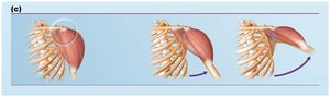
Convergent: Fascicles converge toward a single tendon (e.g., pectoralis major).
Parallel: Fascicles run parallel to the long axis (e.g., sartorius).
Fusiform: Spindle-shaped with parallel fibers (e.g., biceps brachii).
Pennate: Short fascicles attach obliquely to a central tendon. Types include:
Unipennate: Fascicles on one side (e.g., extensor digitorum longus).
Bipennate: Fascicles on both sides (e.g., rectus femoris).
Multipennate: Multiple feather-like fascicles (e.g., deltoid).
Major Muscle Groups and Examples
Some muscles are grouped based on their anatomical or functional relationships:
Epicranius: Frontal belly, Occipital belly
Triceps surae: Gastrocnemius, Soleus
Iliopsoas: Iliacus, Psoas major
Quadriceps femoris: Rectus femoris, Vastus lateralis, Vastus medialis, Vastus intermedius
Hamstrings: Biceps femoris, Semitendinosus, Semimembranosus
Muscles of the Shoulder and Arm
The shoulder and arm contain several important muscles responsible for a wide range of movements:
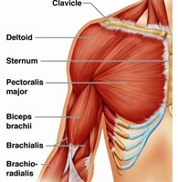
Deltoid: Prime mover of arm abduction.
Pectoralis major: Adduction, medial rotation, and flexion of the arm.
Rotator Cuff Muscles: Supraspinatus, Infraspinatus, Teres minor, Subscapularis (SITS). These muscles stabilize the shoulder joint by keeping the humeral head centered.
Muscles of Respiration
Muscles of the thorax play a key role in breathing:
External intercostals: Elevate the ribs and expand the chest cavity (inspiration).
Internal intercostals: Depress the ribs and decrease chest volume (forced expiration).
Fiber Direction: External intercostals run toward the midline (like hands in pockets); internal intercostals run away from the midline (like crossing arms).
Muscles of the Abdominal Wall
The abdominal wall muscles are involved in trunk movement and stability:
Internal oblique: Rotates torso to the same side.
External oblique: Rotates torso to the opposite side.
Example: To turn the torso left, use the left internal oblique and right external oblique.
Muscles of the Forearm and Wrist
These muscles control movements of the wrist, hand, and fingers. They are organized into anterior (flexor) and posterior (extensor) compartments.
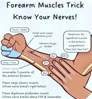
Example: Flexor carpi ulnaris is innervated by the ulnar nerve; flexor digitorum profundus is innervated by both the median and ulnar nerves.
Muscles of the Thigh and Leg
Major muscle groups of the thigh and leg include:
Quadriceps: Rectus femoris, Vastus lateralis, Vastus medialis, Vastus intermedius (anterior thigh; extend the knee).
Hamstrings: Biceps femoris, Semitendinosus, Semimembranosus (posterior thigh; flex the knee).
Triceps surae: Gastrocnemius, Soleus (posterior leg; plantarflex the foot).
Summary Table: Muscle Groupings and Actions
Muscle Group | Muscles Included | Primary Action |
|---|---|---|
Quadriceps femoris | Rectus femoris, Vastus lateralis, Vastus medialis, Vastus intermedius | Knee extension |
Hamstrings | Biceps femoris, Semitendinosus, Semimembranosus | Knee flexion |
Triceps surae | Gastrocnemius, Soleus | Plantarflexion of foot |
Rotator cuff | Supraspinatus, Infraspinatus, Teres minor, Subscapularis | Stabilize shoulder joint |
Key Equations and Concepts
Muscle force production: The force a muscle produces is proportional to its cross-sectional area.
Lever systems: Muscles act on bones as levers to produce movement. The arrangement of the fulcrum, load, and effort determines the type of lever (first, second, or third class).
Additional info: Lever systems in the body are mostly third-class, where the effort is applied between the fulcrum and the load (e.g., biceps brachii flexing the forearm).
