 Back
BackGross Anatomy of the Nervous System: Central and Peripheral Structures
Study Guide - Smart Notes
 Tailored notes based on your materials, expanded with key definitions, examples, and context.
Organization of the Nervous System
Major Subdivisions
The nervous system is divided into two primary parts: the central nervous system (CNS) and the peripheral nervous system (PNS). The CNS includes the brain and spinal cord, while the PNS consists of nerves outside the CNS, further subdivided into cranial and spinal nerves.
CNS: Brain and spinal cord; responsible for processing and integrating information.
PNS: Cranial nerves (originate from the brain) and spinal nerves (originate from the spinal cord); responsible for transmitting information to and from the CNS.
Functional Divisions: Sensory (afferent) neurons bring information to the CNS; motor (efferent) neurons send commands from the CNS to effectors.
Somatic Nervous System (SNS): Controls voluntary skeletal muscle movements.
Autonomic Nervous System (ANS): Controls involuntary functions (heart, glands, viscera); divided into sympathetic and parasympathetic branches.
Autonomic Nervous System (ANS)
The ANS regulates involuntary responses and is divided into two antagonistic branches:
Sympathetic Division: "Fight, flight, freeze"; increases energy expenditure, responds to stress.
Parasympathetic Division: "Rest and digest"; conserves and restores energy.
Autonomic Tone: Both divisions are active to varying degrees, maintaining homeostasis in different organs.
Spinal Cord, Spinal Nerves, and Meninges
Spinal Cord Structure and Function
The spinal cord is the main pathway for information between the brain and body. It can also process some information independently (reflexes).
White Matter: Outer layer; high-speed conduction of nerve impulses.
Grey Matter: Inner "H"-shaped region; information processing, contains neuronal cell bodies.
Central Canal: Filled with cerebrospinal fluid (CSF), continuous with brain cavities.
Spinal Nerves: 31 pairs; each nerve splits into anterior (motor) and posterior (sensory) roots. The posterior root contains the spinal (dorsal root) ganglion.
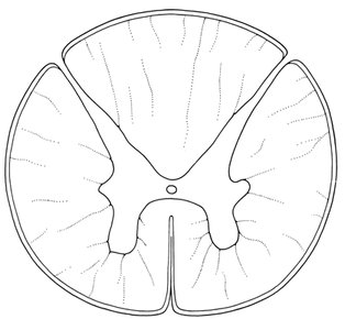
Figure: Transverse cross section of the spinal cord, showing the arrangement of white and grey matter.
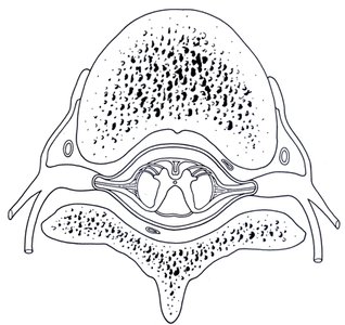
Figure: Cross section of spinal cord with associated nerves and vertebrae, illustrating the anatomical relationship between nervous and skeletal structures.
Meninges and Associated Spaces
The brain and spinal cord are protected by three layers called meninges:
Dura Mater: Outermost, dense irregular connective tissue; in the spinal cord, separated from vertebrae by the epidural space (contains connective, vascular, and adipose tissue).
Arachnoid Mater: Middle layer, thinner and more elastic; separated from dura mater by the subdural space.
Pia Mater: Innermost, highly vascularized, adheres to CNS surfaces; separated from arachnoid mater by the subarachnoid space (contains CSF).
Cerebrospinal Fluid (CSF): Provides shock absorption and maintains CNS environment.
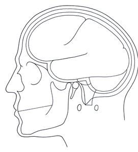
Figure: Sagittal section of the head, illustrating the three meningeal layers and their spatial relationships.
The Brain: Regions and Functions
Major Divisions of the Brain
The brain is divided into four main regions: cerebrum, cerebellum, diencephalon, and brainstem.
Cerebrum: Largest region; divided into left and right hemispheres, further subdivided into frontal, parietal, temporal, and occipital lobes. Responsible for sensory interpretation, motor regulation, emotions, and intellect.
Cerebellum: Posterior to cerebrum; regulates movement, balance, posture, and coordination.
Diencephalon: Includes thalamus (sensory/motor relay), hypothalamus (homeostasis, endocrine control), and pineal gland (melatonin secretion).
Brainstem: Includes midbrain (reflex centers), pons (connects brain regions), and medulla oblongata (regulates cardiovascular and respiratory functions).
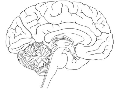
Figure: Sagittal section of the brain, showing the cerebrum, cerebellum, diencephalon, and brainstem.
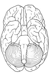
Figure: Inferior view of the brain, highlighting the cerebellum and brainstem.
Main Regions of the Cerebral Cortex and Their Functions
Region | Lobe | Function |
|---|---|---|
Primary Visual (sensory) & Visual Association | Occipital | Vision processing |
Primary Somatosensory, Somatosensory Association, Primary Gustatory | Parietal | Touch, taste, body sensation |
Primary Motor, Motor Speech (production) | Frontal | Voluntary movement, speech |
Primary Auditory (sensory), Auditory Association, Primary Olfactory | Temporal | Hearing, smell |
Diencephalon: Thalamus and Hypothalamus
Thalamus: Relay center for sensory and motor information to the cerebral cortex.
Hypothalamus: Regulates circadian rhythm, sleep-wake cycle, thirst, hunger, body temperature, and controls the pituitary gland.
Pineal Gland: Secretes melatonin, regulates sleep-wake cycle.
Brainstem: Midbrain, Pons, Medulla Oblongata
Midbrain: Pathway for nerve fibers; reflex centers for visual and auditory stimuli.
Pons: Connects medulla oblongata to cerebellum and higher brain centers.
Medulla Oblongata: Regulates cardiovascular and respiratory systems; relay station for sensory information.
Cranial Nerves
Overview and Major Functions
There are twelve cranial nerves, numbered I-XII, each with specific sensory, motor, or mixed functions. Four are studied in detail:
Cranial Nerve | Type | Innervation/Major Function |
|---|---|---|
I Olfactory | Sensory | Nasal mucosa/olfaction |
II Optic | Sensory | Retina/vision |
III Oculomotor | Motor | Eye movement, pupil constriction |
V Trigeminal | Mixed | Facial sensation, chewing |
Olfactory (I): Sense of smell; originates in nasal mucosa, terminates in olfactory bulb and tract.
Optic (II): Vision; originates in retina, passes through optic chiasma, thalamus, and occipital lobe.
Oculomotor (III): Motor control of eye muscles, eyelid, pupil, and lens.
Trigeminal (V): Sensory for face, oral/nasal cavities; motor for chewing and swallowing.
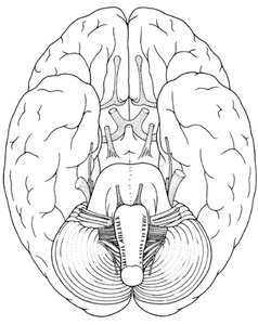
Figure: Inferior view of the brain, showing the cranial nerves and their points of origin.
Clinical Application: Cranial Nerve Neurological Exam
Testing Cranial Nerve Function
Neurological exams assess the function of cranial nerves, often after trauma. Tests include following objects with the eyes (extraocular muscle function), checking facial sensation, and evaluating reflexes. These tests help identify potential nerve damage and guide further medical evaluation.
Summary Table: Nervous System Organization
Division | Main Structures | Function |
|---|---|---|
CNS | Brain, Spinal Cord | Processing, integration, response |
PNS | Cranial & Spinal Nerves | Transmission of information |
SNS | Skeletal Muscles | Voluntary movement |
ANS | Heart, glands, viscera | Involuntary regulation |
Additional info: The notes above expand on brief points from the lab manual, providing academic context and definitions for key terms, as well as tables summarizing major functions and divisions. All included images directly reinforce anatomical explanations and are relevant to the adjacent content.
