 Back
BackGuided Study: Labeling Bones and Structures of the Human Skeleton
Study Guide - Smart Notes
 Tailored notes based on your materials, expanded with key definitions, examples, and context.
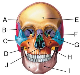
Q1. Label the bones of the skull in the anterior view (A–J).
Background
Topic: Skull Anatomy (Anterior View)
This question tests your ability to identify and label the major bones of the skull from an anterior (front-facing) perspective, which is essential for understanding cranial anatomy in anatomy and physiology (ANP) courses.
Key Terms and Concepts:
Parietal bone: Forms the upper sides and roof of the cranium.
Temporal bone: Located at the sides and base of the skull.
Zygomatic bone: Also known as the cheekbone.
Maxilla bone: Upper jaw bone, holds upper teeth.
Frontal bone: Forehead region.
Sphenoid bone: Butterfly-shaped bone at the base of the skull.
Nasal bone: Bridge of the nose.
Inferior nasal concha: Thin, curved bones in the nasal cavity.
Mandible bone: Lower jaw bone.
Vomer bone: Forms part of the nasal septum.
Step-by-Step Guidance
Start by identifying the most prominent bones in the anterior view: the frontal bone (forehead), maxilla (upper jaw), and mandible (lower jaw).
Locate the zygomatic bones (cheekbones) on either side of the maxilla, and the nasal bones at the bridge of the nose.
Find the parietal and temporal bones at the upper sides and lateral aspects of the skull, respectively.
Identify the sphenoid bone (often visible near the temples or eye sockets), inferior nasal concha (inside the nasal cavity), and vomer bone (midline of the nasal cavity).
Try solving on your own before revealing the answer!
Final Answer:
A. Parietal bone
B. Temporal bone
C. Zygomatic bone
D. Maxilla bone
E. Frontal bone
F. Sphenoid bone
G. Nasal bone
H. Inferior nasal concha
I. Mandible bone
J. Vomer bone
These are the standard anatomical labels for the bones of the skull in the anterior view. Practice locating each on a model or diagram for mastery!
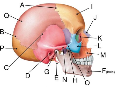
Q2. Label the bones and features of the skull in the lateral view (A–Q).
Background
Topic: Skull Anatomy (Lateral View)
This question assesses your ability to identify the bones and key features of the skull from a side (lateral) perspective, which is crucial for understanding cranial structure and articulation.
Key Terms and Concepts:
Coronal suture: Junction between frontal and parietal bones.
Lambdoid suture: Junction between parietal and occipital bones.
Squamous suture: Junction between parietal and temporal bones.
External acoustic meatus: Ear canal opening.
Styloid process: Pointed projection from the temporal bone.
Mental foramen: Opening in the mandible for nerves/vessels.
Condylar process: Part of mandible that articulates with the skull.
Coronoid process: Anterior projection of the mandible.
Nasal, lacrimal, zygomatic, maxilla, occipital, parietal, temporal bones: Key cranial and facial bones.
Step-by-Step Guidance
Begin by identifying the major sutures (coronal, lambdoid, squamous) and the large bones (parietal, temporal, occipital, frontal).
Locate the facial bones: zygomatic, maxilla, nasal, and lacrimal bones.
Find the processes and foramina: styloid process, external acoustic meatus, mental foramen, condylar and coronoid processes.
Match each label (A–Q) to the corresponding anatomical structure using the key provided.
Try solving on your own before revealing the answer!
Final Answer:
A. Coronal suture
B. Lambdoid suture
C. Squamous suture
D. External acoustic meatus
E. Styloid process of the temporal bone
F. Mental foramen
G. Condylar process of mandible
H. Coronoid process of mandible
J. Nasal bone
K. Lacrimal bone
L. Zygomatic bone
M. Maxilla bone
N. Zygomatic process of the temporal bone
O. Temporal process of the zygomatic bone
P. Occipital bone
Q. Parietal bone
Review the lateral skull diagram to reinforce your understanding of these anatomical landmarks.
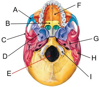
Q3. Label the bones and features of the skull base (inferior view, A–I).
Background
Topic: Skull Base Anatomy (Inferior View)
This question focuses on identifying the bones and foramina visible from the base of the skull, which is important for understanding cranial nerve passage and articulation with the vertebral column.
Key Terms and Concepts:
Zygomatic bone: Cheekbone, visible from below.
Palatine bone: Forms part of the hard palate.
Zygomatic process of the temporal bone: Extension connecting zygomatic and temporal bones.
Sphenoid bone: Central bone at the base of the skull.
Foramen magnum: Large opening for the spinal cord.
Maxilla bone: Upper jaw, forms part of the palate.
Styloid process: Pointed projection from the temporal bone.
Mastoid process: Rounded projection behind the ear.
Occipital condyle: Articulates with the first cervical vertebra.
Step-by-Step Guidance
Identify the central foramen magnum and the occipital condyles on either side.
Locate the maxilla and palatine bones forming the hard palate at the front.
Find the zygomatic bones and their processes, as well as the sphenoid bone near the center.
Spot the styloid and mastoid processes of the temporal bone.
Try solving on your own before revealing the answer!
Final Answer:
A. Zygomatic bone
B. Palatine bone
C. Zygomatic process of the temporal bone
D. Sphenoid bone
E. Foramen magnum
F. Maxilla bone
G. Styloid process of the temporal bone
H. Mastoid process of the temporal bone
I. Occipital condyle
Practice identifying these features on a model or diagram for better retention.
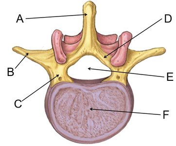
Q4. Label the features of a typical vertebra (A–F).
Background
Topic: Vertebral Anatomy
This question tests your knowledge of the main anatomical features of a typical vertebra, which is foundational for understanding the vertebral column's structure and function.
Key Terms and Concepts:
Spinous process: Posterior projection for muscle/ligament attachment.
Transverse process: Lateral projections for muscle/ligament attachment.
Pedicle: Connects the body to the arch.
Lamina: Part of the vertebral arch.
Vertebral foramen: Opening for the spinal cord.
Body: Main weight-bearing region.
Step-by-Step Guidance
Identify the large, round body at the anterior side of the vertebra.
Locate the vertebral foramen (central hole) and the spinous process (posterior projection).
Find the transverse processes (lateral projections) and the pedicles connecting the body to the arch.
Locate the lamina, which forms part of the vertebral arch.
Try solving on your own before revealing the answer!
Final Answer:
A. Spinous process
B. Transverse process
C. Pedicle
D. Lamina
E. Vertebral foramen
F. Body
Understanding vertebral anatomy is essential for studying the spinal column and its functions.
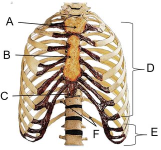
Q5. Label the bones and features of the thoracic cage (A–F).
Background
Topic: Thoracic Cage Anatomy
This question focuses on identifying the bones and features of the thoracic cage, which protects vital organs and supports respiration.
Key Terms and Concepts:
Manubrium: Upper part of the sternum.
Body of the sternum: Central part of the sternum.
Xiphoid process: Inferior tip of the sternum.
True ribs: Ribs directly attached to the sternum.
False ribs: Ribs not directly attached to the sternum.
Floating ribs: Subset of false ribs with no anterior attachment.
Step-by-Step Guidance
Identify the sternum and its three parts: manubrium (top), body (middle), and xiphoid process (bottom).
Locate the true ribs (first seven pairs, directly attached to the sternum).
Find the false ribs (ribs 8–12, not directly attached to the sternum) and the floating ribs (last two pairs, no anterior attachment).
Match each label (A–F) to the corresponding anatomical structure using the key provided.
Try solving on your own before revealing the answer!
Final Answer:
A. Manubrium of the sternum
B. Body of the sternum
C. Xiphoid process of the sternum
D. True ribs
E. False ribs
F. Floating ribs (included as false ribs)
Review the thoracic cage diagram to reinforce your understanding of rib and sternum anatomy.
