 Back
BackGuided Study: Structural and Functional Organization of the Nervous System
Study Guide - Smart Notes
 Tailored notes based on your materials, expanded with key definitions, examples, and context.
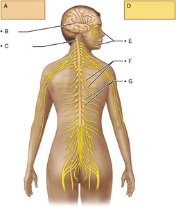
Q5. Name the two principal parts of the nervous system (A and D) and the structures that make up each part (B–C and E–G). Refer to Figure 11.2 for help.
Background
Topic: Structural Organization of the Nervous System
This question tests your understanding of the anatomical divisions of the nervous system and the major structures that belong to each division.
Key Terms:
Central Nervous System (CNS): The main control center, including the brain and spinal cord.
Peripheral Nervous System (PNS): The network of nerves outside the CNS, connecting it to the rest of the body.
Step-by-Step Guidance
Examine the diagram carefully. Identify the two main labeled boxes (A and D) that represent the principal parts of the nervous system.
Recall that the nervous system is divided into the CNS and PNS. The CNS includes the brain and spinal cord, while the PNS includes nerves and ganglia outside the CNS.
For each principal part, list the structures indicated by the labels (B, C for A; E, F, G for D). Use the diagram to match each label to the correct anatomical structure.
Think about the function and location of each structure. For example, the brain and spinal cord are part of the CNS, while nerves branching out from the spinal cord are part of the PNS.
Try solving on your own before revealing the answer!
Final Answer:
A. Central Nervous System (CNS): B. Brain, C. Spinal cord D. Peripheral Nervous System (PNS): E. Cranial nerves, F. Spinal nerves, G. Ganglia
The CNS consists of the brain and spinal cord, while the PNS includes nerves and ganglia that connect the CNS to the rest of the body.
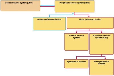
Q6. Use Figure 11.3 to help you complete the flowchart of the organization of the nervous system.
Background
Topic: Functional Organization of the Nervous System
This question is about understanding how the nervous system is organized into divisions and subdivisions, including sensory and motor pathways.
Key Terms:
Sensory (afferent) division: Carries information from receptors to the CNS.
Motor (efferent) division: Carries instructions from the CNS to effectors (muscles and glands).
Somatic nervous system: Controls voluntary movements.
Autonomic nervous system (ANS): Controls involuntary functions.
Sympathetic and parasympathetic divisions: Subdivisions of the ANS.
Step-by-Step Guidance
Look at the flowchart and identify the main branches: CNS and PNS.
Follow the PNS branch to see its subdivisions: sensory (afferent) and motor (efferent).
Trace the motor division to its further subdivisions: somatic and autonomic nervous systems.
Within the autonomic nervous system, identify the sympathetic and parasympathetic divisions.
Try solving on your own before revealing the answer!
Final Answer:
The flowchart should show: Central Nervous System (CNS) and Peripheral Nervous System (PNS); PNS divides into Sensory (afferent) and Motor (efferent); Motor divides into Somatic and Autonomic; Autonomic divides into Sympathetic and Parasympathetic.
This organization helps clarify how information is received, processed, and responded to in the body.
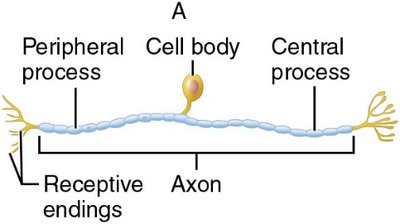
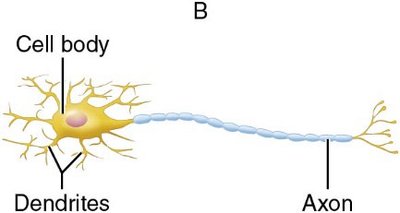
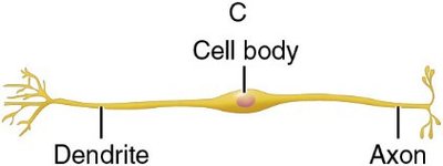
Q10. Assign the appropriate structural classification to each of the neurons (A–C) shown below.
Background
Topic: Structural Classification of Neurons
This question tests your ability to recognize and classify neurons based on their structure: unipolar, bipolar, and multipolar.
Key Terms:
Unipolar neuron: Has one process extending from the cell body.
Bipolar neuron: Has two processes (one axon, one dendrite).
Multipolar neuron: Has many dendrites and one axon.
Step-by-Step Guidance
Examine each neuron diagram (A, B, C) and note the number and arrangement of processes extending from the cell body.
Compare the features of each neuron to the definitions of unipolar, bipolar, and multipolar neurons.
Match each diagram to its structural classification based on the arrangement of dendrites and axons.
Try solving on your own before revealing the answer!
Final Answer:
A. Unipolar neuron B. Multipolar neuron C. Bipolar neuron
Each neuron type is distinguished by the number and arrangement of its processes.
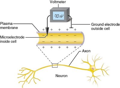
Q8. The voltmeter in the figure below shows a value of −70 mV when measuring the resting membrane potential along the axon of a neuron. What does the minus sign indicate?
Background
Topic: Resting Membrane Potential
This question is about understanding the meaning of the negative value for membrane potential in neurons.
Key Terms:
Resting membrane potential: The voltage difference across the plasma membrane when the cell is at rest.
Millivolts (mV): Unit of electrical potential.
Step-by-Step Guidance
Observe the voltmeter reading and note the value: −70 mV.
Recall that the minus sign indicates the inside of the cell is more negative compared to the outside.
Think about the distribution of ions across the membrane and how this creates the voltage difference.
Try solving on your own before revealing the answer!
Final Answer:
The minus sign indicates that the cytoplasmic side of the plasma membrane is negatively charged relative to the outside.
This is due to the unequal distribution of ions, especially potassium and sodium, across the membrane.
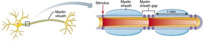
Q13. What is the importance of the myelin sheath gaps (shown below) in saltatory conduction?
Background
Topic: Saltatory Conduction
This question is about understanding how myelin sheath gaps (nodes of Ranvier) facilitate rapid transmission of action potentials along myelinated axons.
Key Terms:
Myelin sheath: Insulating layer around axons.
Myelin sheath gap (node of Ranvier): Exposed region of axon between myelin segments.
Saltatory conduction: The jumping of action potentials from node to node.
Step-by-Step Guidance
Examine the diagram and identify the myelin sheath gaps.
Recall that action potentials are regenerated only at these gaps, not along the entire axon.
Think about how this arrangement increases the speed and efficiency of nerve impulse transmission.
Try solving on your own before revealing the answer!
Final Answer:
Myelin sheath gaps allow action potentials to jump from node to node, greatly increasing conduction velocity and efficiency.
This process is called saltatory conduction and is essential for rapid communication in the nervous system.
