 Back
BackHead and Neck: Skull Bones, Cranial Nerves, and Associated Structures
Study Guide - Smart Notes
 Tailored notes based on your materials, expanded with key definitions, examples, and context.
Head and Neck Anatomy
Overview of Topics
This unit covers the anatomical structures of the head and neck, focusing on the skull bones, cranial nerves, special sense organs, glands, muscles, pharynx, larynx, and neurovasculature. Understanding these components is essential for comprehending the functional organization and clinical relevance of the head and neck region.
Skull bones: Roofing, cranial base, and facial bones
Cranial nerves and brain
Face, oral cavity, nasal cavity
Organs of special senses
Glands
Neck muscles
Pharynx and larynx
Neurovasculature of head and neck
Skull Bones
Classification of Skull Bones
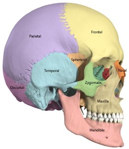
The skull is composed of several bones that protect the brain and form the structure of the face. These bones are classified into three main groups:
Roofing bones: Frontal, Parietal (2)
Cranial base bones: Ethmoid, Sphenoid, Temporal (2), Occipital
Facial bones: Maxilla, Zygomatic, Nasal, Lacrimal, Inferior nasal concha, Vomer, Mandible, Palatine
Major Sutures and Fontanelles
Sutures are immovable joints between skull bones, while fontanelles are soft spots in the fetal and infant skull that allow for growth.
Sutures: Sagittal, Coronal, Squamosal, Lambdoidal
Fontanelles: Anterior (closes 18–24 months), Posterior (2–3 months), Sphenoidal (6 months), Mastoid (6–18 months)
Cranial Base Bones
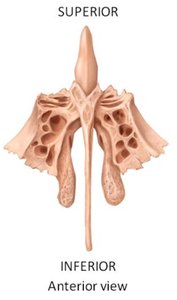
Ethmoid Bone
The ethmoid bone forms part of the anterior cranial base and the nasal cavity. It contains the crista galli, perpendicular plate, cribriform plate, and nasal conchae.
Crista galli: Attachment for the falx cerebri
Cribriform plate: Passage for olfactory nerves (CN I)
Superior and middle nasal conchae: Increase surface area in the nasal cavity
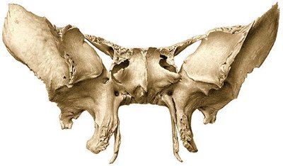
Sphenoid Bone
The sphenoid bone is a central bone of the cranial base, contributing to the floor of the cranium and the orbits. It contains the body, lesser and greater wings, pterygoid processes, and several foramina for cranial nerves.
Optic canal: Passage for optic nerve (CN II)
Superior orbital fissure: Passage for CN III, IV, V1, VI
Foramen rotundum: Passage for maxillary nerve (CN V2)
Foramen ovale: Passage for mandibular nerve (CN V3)
Hypophyseal fossa: Houses the pituitary gland
Temporal Bone
The temporal bone houses structures for hearing and balance and forms part of the cranial base and lateral skull.
Regions: Squamous, tympanic, mastoid, petrous
Key features: External and internal acoustic meatus, zygomatic process, styloid process
Foramina: Passage for facial (CN VII) and vestibulocochlear (CN VIII) nerves
Occipital Bone
The occipital bone forms the posterior part of the cranial base and contains the foramen magnum for the spinal cord.
Regions: Squamous, basilar
Foramen magnum: Passage for medulla oblongata and vertebral arteries
Hypoglossal canal: Passage for hypoglossal nerve (CN XII)
Jugular foramen: Passage for CN IX, X, XI and internal jugular vein
Carotid canal: Passage for internal carotid artery
Cranial Nerves and Foramina
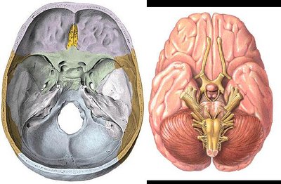
Overview of Cranial Nerves
The cranial nerves exit the cranium through foramina in the cranial base bones. Each nerve has a specific function, including sensory, motor, or both.
CN I: Olfactory (smell)
CN II: Optic (vision)
CN III: Oculomotor (eye movement)
CN IV: Trochlear (eye movement)
CN V: Trigeminal (facial sensation, mastication)
CN VI: Abducens (eye movement)
CN VII: Facial (facial expression, taste)
CN VIII: Vestibulocochlear (hearing, balance)
CN IX: Glossopharyngeal (taste, swallowing)
CN X: Vagus (parasympathetic control)
CN XI: Spinal accessory (neck muscles)
CN XII: Hypoglossal (tongue movement)
Key Foramina and Their Contents
Foramen | Bone | Contents |
|---|---|---|
Cribriform plate | Ethmoid | Olfactory nerves (CN I) |
Optic canal | Sphenoid | Optic nerve (CN II) |
Superior orbital fissure | Sphenoid | CN III, IV, V1, VI |
Foramen rotundum | Sphenoid | Maxillary nerve (CN V2) |
Foramen ovale | Sphenoid | Mandibular nerve (CN V3) |
Internal acoustic meatus | Temporal | CN VII, VIII |
Jugular foramen | Occipital/Temporal | CN IX, X, XI; Internal jugular vein |
Hypoglossal canal | Occipital | Hypoglossal nerve (CN XII) |
Carotid canal | Temporal | Internal carotid artery |
Facial Bones
Major Facial Bones and Their Functions
The facial bones form the structure of the face, support the teeth, and contribute to the orbits and nasal cavity.
Maxilla: Upper jaw, forms part of the hard palate
Zygomatic: Cheekbone
Nasal: Bridge of the nose
Lacrimal: Medial wall of orbit
Inferior nasal concha: Lateral wall of nasal cavity
Vomer: Inferior part of nasal septum
Mandible: Lower jaw, only movable skull bone
Palatine: Posterior part of hard palate
Additional Info
The skull bones are joined by sutures, which ossify with age.
Fontanelles allow for brain growth and facilitate childbirth.
Special sense organs (smell, vision, hearing, balance) are closely associated with cranial base bones and their foramina.
