 Back
BackHearing: Anatomy and Physiology of the Ear and Signal Transduction
Study Guide - Smart Notes
 Tailored notes based on your materials, expanded with key definitions, examples, and context.
Hearing and the Anatomy of the Ear
Structure of the Ear
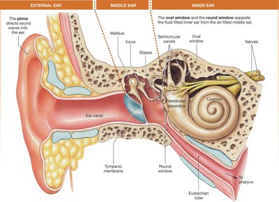
The human ear is divided into three main regions: the external ear, middle ear, and inner ear. Each region plays a distinct role in the process of hearing, from capturing sound waves to converting them into electrical signals for the brain.
External Ear: Includes the pinna (outer ear), which directs sound waves into the ear canal, and the ear canal, which conducts sound waves to the tympanic membrane.
Middle Ear: Contains three small bones—the malleus, incus, and stapes—that transfer vibrations from the tympanic membrane to the inner ear.
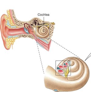
Inner Ear: Houses sensory structures for equilibrium (vestibular apparatus, semicircular canals) and hearing (cochlea, oval window, round window).
Features of Sound Waves
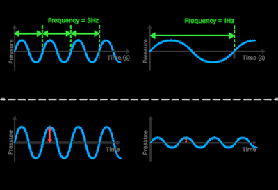
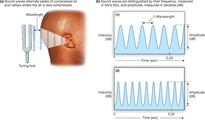
Sound waves are mechanical vibrations that travel through air and are interpreted by the central nervous system (CNS) as hearing. The two main features of sound waves are frequency and amplitude.
Frequency: Determines the pitch of the sound and is measured in hertz (Hz). Higher frequency corresponds to higher pitch.
Amplitude: Determines the loudness of the sound and is measured in decibels (dB). Greater amplitude means louder sound.
Signal Transduction: From Sound Wave to Electrical Signal
Five Steps of Signal Transduction
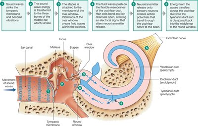
The process of hearing involves five key steps that convert sound waves into electrical signals processed by the brain:
Sound waves strike the tympanic membrane and become vibrations.
The sound wave energy is transferred to the three bones of the middle ear, which vibrate.
The stapes is attached to the membrane of the oval window. Vibrations of the oval window create fluid waves within the cochlea.
Fluid waves push on the basilar membrane of the cochlear duct. Hair cells bend and release neurotransmitter.
Neurotransmitter release onto sensory neurons creates action potentials that travel through the cochlear nerve to the brain.
The Cochlea: Structure and Function
Anatomy of the Cochlea
The cochlea is a spiral-shaped organ in the inner ear responsible for converting mechanical vibrations into neural signals. It contains three parallel, fluid-filled channels:
Vestibular and Tympanic Ducts: Continuous with each other and filled with perilymph (similar to plasma).
Cochlear Duct: A dead-end tube between the vestibular and tympanic ducts, filled with endolymph (similar to intracellular fluid).
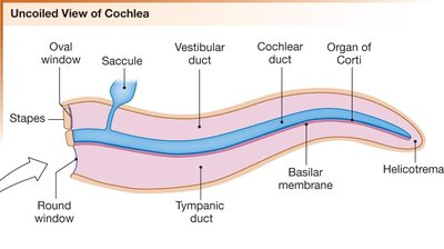
Organ of Corti and Hair Cells
The cochlear duct contains the Organ of Corti, which sits on the basilar membrane and is partially covered by the tectorial membrane. The Organ of Corti houses hair cell receptors and support cells, which are essential for the transduction of sound.
Hair Cells: Sensory cells that detect movement of the basilar membrane and convert it into electrical signals.
Basilar Membrane: Vibrates in response to fluid waves, causing hair cells to bend.
Tectorial Membrane: Covers the hair cells and plays a role in their activation.
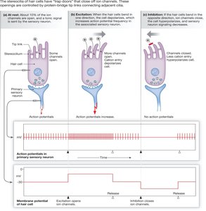
Mechanism of Hair Cell Activation
Bending of Stereocilia and Neurotransmitter Release
When the basilar membrane moves relative to the tectorial membrane, the stereocilia (hair-like projections) on the auditory hair cells bend. This bending opens or closes ion channels, leading to changes in membrane potential and neurotransmitter release.
Bending Toward Tallest Stereocilia: Opens ion channels, increases neurotransmitter release, and enhances action potential frequency in sensory neurons.
Bending Away from Tallest Stereocilia: Closes ion channels, decreases neurotransmitter release, and reduces action potential frequency.
Summary Table: Steps in Auditory Signal Transduction
The following table summarizes the main steps in the conversion of sound waves to electrical signals:
Step | Description |
|---|---|
1 | Sound waves strike tympanic membrane |
2 | Middle ear bones vibrate |
3 | Stapes vibrates oval window, creating fluid waves in cochlea |
4 | Fluid waves move basilar membrane, bending hair cells |
5 | Neurotransmitter release triggers action potentials in cochlear nerve |
Key Terms and Concepts
Pinna: Outer ear structure that directs sound waves.
Tympanic Membrane: Eardrum; separates external and middle ear.
Malleus, Incus, Stapes: Middle ear bones that transmit vibrations.
Cochlea: Inner ear organ for hearing.
Organ of Corti: Structure containing hair cells for sound transduction.
Stereocilia: Hair-like projections on hair cells that bend in response to sound.
Perilymph/Endolymph: Fluids in cochlear ducts.
Basilar Membrane: Vibrates in response to sound, activating hair cells.
Tectorial Membrane: Covers hair cells in the cochlea.
Equations and Measurements
Frequency (Pitch): (measured in Hz)
Amplitude (Loudness): (measured in dB)
Example: Application in Hearing
When a tuning fork is struck, it produces sound waves that travel through the air. The pinna directs these waves into the ear canal, where they strike the tympanic membrane. Vibrations are transferred through the middle ear bones to the cochlea, where fluid waves activate hair cells. The resulting electrical signals are sent to the brain, allowing us to perceive the sound.
Additional info:
The vestibular apparatus and semicircular canals in the inner ear are responsible for equilibrium, not hearing.
Hair cells are specialized mechanoreceptors that convert mechanical energy into neural signals.
